MedicosNotes.com

A site for medical students - Practical,Theory,Osce Notes

>

Exploring Radiofemoral Delay: Understanding Its Mechanisms and Identifying Its Causes


What is Radiofemoral Delay and What are its Causes?

Radiofemoral delay is a clinical sign indicative of a significant delay between the palpation of the radial pulse (at the wrist) and the femoral pulse (in the groin). This phenomenon is often associated with specific cardiovascular conditions and can be a critical clue in diagnosing vascular diseases. Understanding the implications and causes of radiofemoral delay is essential for healthcare professionals, as it can guide further diagnostic evaluations and management strategies.

Understanding the Circulatory Pathway

To comprehend radiofemoral delay, it's crucial to have a basic understanding of the body's circulatory system. Blood is pumped from the heart through the arteries, delivering oxygen and nutrients to various body tissues. The radial artery in the wrist and the femoral artery in the groin are both key components of this arterial system, supplying blood to the lower and upper limbs, respectively.

Mechanism Behind Radiofemoral Delay

Under normal circumstances, the pulse waves generated by the heartbeat are transmitted simultaneously through the aorta and its branches, reaching the radial and femoral arteries almost at the same time. Therefore, in a healthy individual, there should be no noticeable delay when palpating these pulses sequentially.

Radiofemoral delay occurs when there is a disruption or obstruction in the blood flow from the heart towards the lower part of the body, specifically affecting the aorta's ability to efficiently deliver blood to the femoral artery. This disruption results in a noticeable delay in the pulse wave reaching the femoral artery compared to the radial artery.

Causes of Radiofemoral Delay

The causes of radiofemoral delay can generally be categorized into congenital (present at birth) and acquired conditions that affect the aorta or its major branches. Some of the most common causes include:

  1. Coarctation of the Aorta (CoA): A congenital condition characterized by a narrowing of a section of the aorta. This narrowing can obstruct blood flow, leading to a significant delay in the pulse wave reaching the femoral artery compared to the radial artery.

  2. Aortic Dissection: This is a critical condition where there is a tear in the inner layer of the aorta's wall. Blood enters the wall of the artery, creating a new channel and disrupting normal blood flow. This can significantly impact the timing of pulse waves.

  3. Atherosclerosis: The buildup of plaque inside the artery walls can narrow and harden the arteries, reducing blood flow. When atherosclerosis affects the aorta or its branches leading to the lower body, it can cause radiofemoral delay.

  4. Takayasu’s Arteritis: A rare inflammatory disease that damages the aorta and its main branches. The inflammation can lead to narrowing, occlusion, or aneurysm of these arteries, affecting the pulse wave velocity.

  5. Other Vascular Anomalies: Rarely, other vascular conditions, such as aneurysms or arteriovenous malformations (abnormal connections between arteries and veins), can affect the timing and strength of pulse waves, leading to a radiofemoral delay.

Diagnosis and Importance

The detection of radiofemoral delay is usually performed through a physical examination, where a healthcare provider palpates the radial and femoral pulses simultaneously or in quick succession. When a delay is suspected, further diagnostic tests such as Doppler ultrasound, CT angiography, or MRI may be employed to visualize the blood flow and structures of the arteries.

Recognizing radiofemoral delay is crucial as it may be the first clue to underlying serious cardiovascular conditions that require prompt intervention. Early diagnosis and treatment of the underlying cause are vital to prevent complications and improve patient outcomes.

Conclusion

Radiofemoral delay is more than a mere discrepancy in pulse timing; it's a window into the vascular health of an individual. Understanding its causes and implications enables healthcare professionals to undertake timely and appropriate interventions, ultimately safeguarding cardiovascular health.


Newer Targets for Treatment of Asthma: A Glimpse into the Future


Asthma, a chronic respiratory disease characterized by inflammation and narrowing of the airways, affects millions of people worldwide. Despite existing treatment options, asthma remains uncontrolled in a significant proportion of patients, necessitating research into novel therapeutic targets. This article explores some of the promising new targets currently being investigated for the treatment of asthma.

Biologic Therapies

Biologic therapies, which target specific molecules involved in the immune response, have emerged as a promising area of asthma treatment.

  1. Anti-Interleukin-5 (IL-5) and Anti-IL-5R Therapies: IL-5 plays a key role in the maturation and survival of eosinophils, a type of white blood cell involved in asthma inflammation. Biologics such as mepolizumab, reslizumab, and benralizumab target IL-5 or its receptor (IL-5R), reducing eosinophilic inflammation and the frequency of asthma exacerbations.
  2. Anti-Interleukin-4 (IL-4) and Anti-Interleukin-13 (IL-13) Therapies: IL-4 and IL-13 are also crucial in the immune response leading to asthma. Dupilumab, a biologic drug that inhibits both IL-4 and IL-13, has shown promise in the treatment of moderate-to-severe asthma.

Bronchial Thermoplasty

Bronchial thermoplasty is a novel non-pharmacological intervention for severe asthma. It involves applying controlled thermal energy to the airway walls during a series of bronchoscopy procedures, reducing the amount of airway smooth muscle and thereby diminishing the airways' ability to constrict.

Targeting Neutrophilic Asthma

While eosinophilic asthma has been the focus of many new therapies, neutrophilic asthma, another subtype of the disease, has proven more challenging. However, new targets are being explored:

Anti-Interleukin-17 (IL-17) Therapy: IL-17 has been associated with neutrophilic inflammation in asthma. Anti-IL-17 therapies are being investigated for their potential to reduce neutrophilic airway inflammation.

Anti-Interleukin-8 (IL-8) Therapy: As a potent neutrophil attractant, IL-8 is another potential target in neutrophilic asthma. Research is ongoing to develop therapies that can block IL-8 or its receptor.

Phosphodiesterase-4 (PDE4) Inhibitors: PDE4 inhibitors, such as roflumilast, can reduce inflammation and are being investigated for use in severe neutrophilic asthma.

Emerging Targets

Other potential treatment targets include toll-like receptors (TLRs), which play a role in the immune response, and chitinase-like proteins (CLPs), associated with inflammation and tissue remodeling in asthma.

The landscape of asthma treatment is evolving, with promising new therapies targeting the underlying pathophysiology of the disease. As our understanding of asthma's complex immunological and physiological processes deepens, we can expect even more sophisticated and effective treatments to emerge, offering hope for those living with this chronic condition. However, as with any new therapeutic strategies, these potential treatments must undergo rigorous testing for safety and efficacy before they can be incorporated into routine clinical practice.

Assessment of Patient Fitness for Thoracic Surgery

Thoracic surgery, involving procedures on the lungs, heart, and other structures within the chest cavity, is a major intervention that can pose significant risks. Hence, preoperative assessment of a patient's fitness for thoracic surgery is crucial to determine surgical feasibility, inform patients of potential risks and benefits, and optimize preoperative conditions for a favorable surgical outcome.

Patient Evaluation

  1. History and Physical Examination: Every preoperative evaluation begins with a comprehensive medical history and physical examination. Factors such as age, smoking history, preexisting conditions (like chronic obstructive pulmonary disease (COPD), asthma, cardiovascular disease, or diabetes), and previous surgeries can significantly influence the risk profile.
  2. Pulmonary Function Testing (PFT): PFT is a key part of preoperative assessment for thoracic surgery. It provides vital information about lung function and can predict postoperative pulmonary function. The forced expiratory volume in one second (FEV1) and diffusion capacity of the lung for carbon monoxide (DLCO) are particularly important measures.
  3. Cardiac Evaluation: Cardiovascular disease is a common comorbidity in patients undergoing thoracic surgery. A thorough cardiac evaluation may include an electrocardiogram (ECG), echocardiography, or stress testing. In specific cases, a coronary angiography may be necessary.
  4. Imaging: CT scans of the chest provide crucial information about the disease's location, extent, and potential surgical approach. They also help identify any unforeseen issues, like unexpected metastasis in cases of lung cancer.
  5. Nutritional Status: Malnutrition can lead to delayed wound healing and increase the risk of postoperative complications. Assessing nutritional status, including parameters like BMI and serum albumin levels, is crucial.
  6. Performance Status: Tools like the Eastern Cooperative Oncology Group (ECOG) or Karnofsky Performance Status (KPS) scales help assess a patient's ability to perform everyday tasks, providing insights into their overall health status and resilience.
  7. Exercise Testing: Exercise tests, such as the six-minute walk test or cardiopulmonary exercise testing (CPET), provide objective measures of a patient's aerobic fitness and endurance.

Risk Stratification and Optimization

Assessment data is used to stratify patients into risk categories. High-risk patients may need additional investigations and interventions to optimize their condition before surgery. Smoking cessation, pulmonary rehabilitation, and optimization of any preexisting conditions (like diabetes or hypertension) are key components of preoperative optimization.

Informed Consent

Once fitness for surgery is established, it is crucial to discuss the potential risks and benefits with the patient. This conversation should encompass not only the surgical risks but also the expected postoperative recovery period and the impact on the patient's quality of life.

Assessing patient fitness for thoracic surgery is a comprehensive process, demanding careful consideration of multiple factors, including pulmonary function, cardiac health, and overall performance status. It enables clinicians to identify potential risks, optimize patient condition before surgery, and set realistic expectations, thereby paving the way for successful surgical outcomes. As each patient is unique, the assessment must be personalized and nuanced, balancing the potential benefits of surgery against its risks.

Understanding the Oxygen Dissociation Curve

The oxygen dissociation curve is a graphical representation that delineates the relationship between the partial pressure of oxygen (pO2) in the blood and the oxygen saturation of hemoglobin, the protein in red blood cells responsible for transporting oxygen. This curve is crucial for understanding how oxygen is delivered to body tissues and cells, and how changes in environmental or physiological conditions affect oxygen transportation and availability.

Understanding Hemoglobin and Oxygen Transport

Hemoglobin is a globular, iron-containing protein within red blood cells, uniquely suited for oxygen binding and transportation. Each hemoglobin molecule can bind up to four oxygen molecules, a process called oxygenation. When oxygen binds to hemoglobin, it forms oxyhemoglobin.

When blood flows through the lungs, oxygen molecules bind to the hemoglobin in red blood cells. This oxygen-rich blood then travels to the body's tissues and organs, where the oxygen dissociates from the hemoglobin (hence the term "oxygen dissociation") and is delivered to cells for their metabolic activities.

The Oxygen Dissociation Curve

The oxygen dissociation curve is typically an S-shaped (sigmoidal) curve. The x-axis represents the partial pressure of oxygen in the blood, and the y-axis represents the percentage of hemoglobin saturated with oxygen. The curve's shape is primarily due to the cooperative binding nature of hemoglobin.

When the blood's pO2 is low, such as in the metabolically active tissues, hemoglobin releases its oxygen (a condition favoring oxygen 'dissociation'). Conversely, when the pO2 is high, as in the lungs, hemoglobin binds to oxygen. This relationship between oxygen's partial pressure and its saturation is the essence of the oxygen dissociation curve.

Factors Affecting the Oxygen Dissociation Curve

Several factors can shift the oxygen dissociation curve to the left or right, altering hemoglobin's affinity for oxygen:

  1. Temperature: An increase in body temperature shifts the curve to the right, indicating decreased oxygen affinity. This situation is often seen during fever or heavy exercise.
  2. pH (Bohr Effect): The Bohr effect states that a decrease in pH (indicating an increase in blood acidity) shifts the curve to the right, again, lowering hemoglobin's affinity for oxygen. This effect allows more oxygen to be released in metabolically active tissues that produce more acidic waste products.
  3. Carbon Dioxide (CO2) Levels: High levels of CO2 also shift the curve to the right. CO2 is a byproduct of cellular metabolism and increases in the blood during vigorous exercise or in certain health conditions.
  4. 2,3-Diphosphoglycerate (2,3-DPG): This compound, produced by red blood cells, reduces hemoglobin's affinity for oxygen. Increased levels of 2,3-DPG, as seen in conditions like anemia or high altitude, shift the curve to the right, facilitating oxygen release to tissues.

The oxygen dissociation curve is an integral tool for understanding oxygen transport and delivery in the body. Its sigmoidal shape reflects the cooperative binding of oxygen to hemoglobin, and shifts in the curve due to various physiological or environmental conditions help ensure that tissues receive the oxygen they need. Understanding these concepts is crucial in fields like physiology, medicine, and biomedical research.

Physiological changes in lung at high altitude and pathophysiology of high altitude pulmonary edema

At high altitudes, where the oxygen concentration in the air is lower, the body undergoes several physiological changes to adapt to the reduced oxygen availability. These changes primarily occur in the lungs and cardiovascular system. Here are some of the key physiological changes that take place:

  1. Hyperventilation: At high altitudes, the body increases its respiratory rate and depth of breathing to compensate for the reduced oxygen levels. This hyperventilation helps maintain adequate oxygen uptake and carbon dioxide elimination.
  2. Increased pulmonary blood pressure: In response to low oxygen levels, the blood vessels in the lungs constrict (pulmonary vasoconstriction), causing an increase in pulmonary blood pressure. This redirection of blood flow helps optimize oxygen delivery to the body's tissues.
  3. Increased red blood cell production: The body responds to high-altitude conditions by producing more red blood cells (erythropoiesis). This increase in red blood cells helps enhance the oxygen-carrying capacity of the blood.
  4. Altered gas exchange: The efficiency of gas exchange in the lungs may be impaired at high altitudes due to factors such as reduced oxygen pressure and increased diffusion distance. However, the body's compensatory mechanisms, including hyperventilation and increased red blood cell production, help mitigate the impact of these changes.

Despite these adaptive mechanisms, some individuals may still develop high altitude pulmonary edema (HAPE), which is a potentially life-threatening condition. HAPE is a type of non-cardiogenic pulmonary edema that occurs at high altitudes and is characterized by the accumulation of fluid in the lungs. The exact pathophysiology of HAPE is not completely understood, but several factors contribute to its development:

  1. Increased pulmonary artery pressure: The constriction of blood vessels in the lungs at high altitudes can lead to increased pulmonary artery pressure. This elevated pressure can cause leakage of fluid from the pulmonary capillaries into the lung tissue.
  2. Increased capillary permeability: The increased pulmonary artery pressure and the hypoxic environment at high altitudes can cause damage to the endothelial lining of the pulmonary capillaries. This damage results in increased capillary permeability, allowing fluid to leak into the alveoli.
  3. Inflammation: Hypoxia and other factors at high altitudes can trigger an inflammatory response in the lungs. Inflammatory mediators and increased vascular permeability further contribute to the accumulation of fluid in the alveoli.
  4. Reduced clearance of fluid: The impaired lymphatic drainage from the lungs at high altitudes can hinder the clearance of fluid, exacerbating the accumulation of fluid in the alveoli.

The accumulation of fluid in the lungs leads to impaired gas exchange, causing symptoms such as shortness of breath, cough, wheezing, and fatigue. If left untreated, HAPE can progress rapidly and result in severe respiratory distress and even respiratory failure.

The management of HAPE involves immediate descent to lower altitudes, administration of supplemental oxygen, and the use of medications such as diuretics to reduce fluid accumulation. Prompt medical attention is crucial for the effective treatment of HAPE.

Normal process of respiration, Oxygen uptake in the blood and elimination of CO2 from the body

Respiration is the process by which living organisms take in oxygen from the environment and release carbon dioxide. In humans, respiration involves two main processes: external respiration and internal respiration.

External respiration occurs in the lungs and involves the exchange of gases between the air in the lungs and the bloodstream. When we inhale, air enters the respiratory system and reaches the alveoli, which are tiny air sacs in the lungs. The alveoli are surrounded by a network of capillaries, where the exchange of gases takes place.

Oxygen (O2) from the inhaled air diffuses across the thin walls of the alveoli and enters the bloodstream. It binds to hemoglobin, a protein in red blood cells, forming oxyhemoglobin. This oxygenated blood is then transported to the body's tissues.

Internal respiration occurs at the tissue level, where oxygen is delivered to cells and carbon dioxide (CO2) is produced as a waste product of cellular metabolism. In the tissues, oxygen detaches from hemoglobin and diffuses into the cells, where it is used in the process of cellular respiration to produce energy.

During cellular respiration, glucose and oxygen react to produce carbon dioxide, water, and energy in the form of adenosine triphosphate (ATP). The carbon dioxide generated as a byproduct diffuses out of the cells into the surrounding capillaries.

The oxygen-depleted blood returns to the heart and is pumped to the lungs through the pulmonary artery. In the lungs, carbon dioxide diffuses from the capillaries into the alveoli, and it is then exhaled out of the body during the process of exhalation.

Oxygen uptake in the blood is facilitated by the high affinity of hemoglobin for oxygen. Hemoglobin binds to oxygen in the lungs, and this binding is reversible, allowing for oxygen to be released in the tissues where it is needed. The oxygen-carrying capacity of blood is influenced by factors such as hemoglobin concentration, blood pH, temperature, and partial pressure of oxygen.

The elimination of carbon dioxide occurs through the lungs during exhalation. Carbon dioxide in the bloodstream combines with water to form carbonic acid (H2CO3), which quickly dissociates into bicarbonate ions (HCO3-) and hydrogen ions (H+). The bicarbonate ions are transported back to the lungs through the bloodstream, where they are converted back into carbon dioxide, which is then exhaled.

Hypoxia refers to a condition in which the body or a region of the body is deprived of adequate oxygen supply. There are various forms of hypoxia, including:

  1. Hypoxic hypoxia: This occurs when there is a decrease in the oxygen concentration in the air, such as at high altitudes or in poorly ventilated environments.
  2. Anemic hypoxia: It results from a decrease in the oxygen-carrying capacity of blood due to a decrease in the number of red blood cells or a decrease in the amount of hemoglobin.
  3. Ischemic hypoxia: It happens when there is a reduction in blood flow to tissues, leading to inadequate oxygen supply. It can be caused by conditions such as circulatory disorders or blockages in blood vessels.
  4. Histotoxic hypoxia: This occurs when the cells are unable to utilize oxygen effectively due to the presence of toxins or metabolic poisons, impairing cellular respiration.
  5. Hypoxemic hypoxia: It results from a decrease in the oxygen content of the blood, usually caused by respiratory disorders, such as lung diseases or impaired gas exchange in the lungs.

These forms of hypoxia can have varying degrees of severity and can lead to symptoms ranging from mild breathlessness and fatigue to more severe complications affecting vital organs. Prompt medical attention and appropriate interventions are necessary to address hypoxic conditions and restore oxygen supply to the body's tissues.

Chemical Carcinogens: An In-depth Overview

Chemical carcinogens are substances that can contribute to the development of cancer by causing changes (mutations) in a cell's DNA or leading to other types of cellular damage. These changes can potentially lead to uncontrolled cell division and growth, the hallmarks of cancer. This article provides a comprehensive overview of chemical carcinogens, their sources, mechanisms of action, and strategies to minimize exposure.

Understanding Chemical Carcinogens

Chemical carcinogens are classified based on their ability to initiate or promote cancer. Initiators are chemicals that cause DNA damage and mutations, while promoters stimulate the proliferation of these mutated cells but do not cause mutations themselves. Many chemical carcinogens are capable of both initiating and promoting cancer.

Sources of Chemical Carcinogens

Chemical carcinogens are found in various sources, including:

  1. Tobacco Smoke: This contains over 60 known carcinogens, including polycyclic aromatic hydrocarbons (PAHs), N-nitrosamines, and aldehydes. These are responsible for the strong association between tobacco use and cancers of the lung, mouth, throat, and other organs.
  2. Diet: Certain foods can contain chemical carcinogens. For instance, aflatoxins produced by molds on improperly stored grains and nuts are potent liver carcinogens. Processed meats often contain N-nitroso compounds, while high-temperature cooking can produce heterocyclic amines and PAHs.
  3. Occupational Exposure: Certain industries expose workers to carcinogens, such as asbestos in construction, benzene in chemical manufacturing, and coal tar in metalworking industries.
  4. Environment: Exposure can occur through polluted air, water, or soil. Common environmental carcinogens include asbestos, arsenic, and certain byproducts of industrial processes.

Mechanisms of Action

Chemical carcinogens can damage DNA directly or require metabolic activation to become carcinogenic. For instance, benzene, a known leukemogen, is metabolized in the liver to produce reactive intermediates, which can cause DNA damage leading to leukemia.

Chemical carcinogens can also cause cancer through non-genotoxic mechanisms, such as inducing chronic inflammation, suppressing immune responses, or disrupting cell signaling pathways that regulate cell growth and differentiation.

Mitigating Exposure to Chemical Carcinogens

Reducing exposure to chemical carcinogens is a key strategy in cancer prevention. This can be achieved through various means:

  1. Lifestyle Choices: Avoiding tobacco, limiting alcohol consumption, eating a healthy diet, and exercising regularly can significantly reduce the risk of cancer.
  2. Occupational Safety: Implementing safety regulations and protective measures in workplaces can minimize exposure to occupational carcinogens.
  3. Environmental Regulations: Enforcing laws to control pollution and limit public exposure to environmental carcinogens is crucial.
  4. Education and Awareness: Public awareness campaigns about the risks of carcinogens can encourage healthier behaviors and demand for safer products.

Pain Management in Patients with Spinal Cord Compression


Spinal cord compression (SCC) is a serious condition that occurs when a mass places pressure on the spinal cord. This pressure can originate from various sources, including a herniated disc, a bone fracture, or a tumor. Patients with SCC often experience significant pain, in addition to other neurological symptoms such as weakness or numbness. Consequently, pain management is a critical aspect of care for these patients. This article explores the various strategies and treatments for pain management in patients with spinal cord compression.

Understanding Spinal Cord Compression Pain

The pain associated with SCC can manifest in several ways. It may present as back or neck pain at the site of the compression, radiating pain that spreads to the limbs, or even as a band-like pain around the trunk. This pain can severely affect a patient's quality of life, making effective pain management strategies crucial.

Non-Pharmacological Interventions

Non-drug approaches are often used as adjuncts to medication in managing pain from SCC. These can include:

  1. Physical Therapy: Specific exercises can help alleviate some types of pain and improve mobility and strength.
  2. Occupational Therapy: This can teach coping strategies and modifications to daily activities to help manage pain and improve function.
  3. Psychological Support: Cognitive-behavioral therapy, relaxation techniques, and other psychological interventions can help patients manage the emotional impact of chronic pain.

Pharmacological Management

Medication is often the first line of treatment for SCC-related pain:

  1. Non-Steroidal Anti-Inflammatory Drugs (NSAIDs): These drugs can help reduce pain and inflammation. They are often used for mild to moderate pain.
  2. Corticosteroids: These can reduce inflammation and swelling around the spinal cord, relieving pressure and pain.
  3. Opioids: For severe pain, opioids may be prescribed. However, their use needs careful monitoring due to the risk of dependency and side effects.
  4. Adjuvant Analgesics: Certain antidepressants and anticonvulsants can help manage neuropathic pain often associated with SCC.

Interventional Techniques

When medication and non-drug interventions are insufficient, more invasive strategies may be considered:

  1. Nerve Blocks: These involve injecting medication around specific nerves or into the epidural space to block pain signals.
  2. Neurostimulation Devices: These devices deliver electrical stimulation to the spinal cord or specific nerves to block the perception of pain.
  3. Intrathecal Pumps: These devices deliver pain medication directly to the space around the spinal cord.

Surgical Intervention

In some cases, surgery may be required to remove or reduce the source of the compression. While the primary goal is to alleviate the pressure on the spinal cord, surgery can also significantly reduce pain.

Palliative Care

For patients with advanced disease where the focus is on comfort rather than cure, palliative care plays an essential role. This approach prioritizes quality of life, symptom relief, and psychosocial support.

Pain management in patients with spinal cord compression involves a multifaceted approach, considering non-pharmacological methods, medications, interventional techniques, possible surgical intervention, and palliative care. Ultimately, the goal is to improve the patient's quality of life by effectively managing pain and enhancing overall functionality. Given the complexity of SCC and its associated pain, a personalized, multidisciplinary approach is key to successful management.

Role of Surgery in Abdominal Non-Hodgkin’s Lymphoma


Non-Hodgkin’s lymphoma (NHL) is a heterogeneous group of malignancies of the lymphatic system. While the disease primarily involves lymph nodes, it can also arise in extranodal sites, with the gastrointestinal (GI) tract being the most commonly affected site. Abdominal non-Hodgkin’s lymphoma can involve any part of the GI tract, from the stomach and small intestine to the colon and rectum. The role of surgery in the management of abdominal NHL has evolved significantly over the years and remains a topic of ongoing debate. This article reviews the current understanding and application of surgery in the treatment of abdominal non-Hodgkin’s lymphoma.

Therapeutic Paradigms and the Role of Surgery

Traditionally, the primary modality for treating non-Hodgkin's lymphoma has been chemotherapy, with or without radiotherapy, based on the type and stage of the disease. However, the role of surgery has shifted from a therapeutic to a largely diagnostic and supportive role.

Today, surgical intervention in abdominal NHL is generally reserved for specific situations, including:

  1. Diagnosis and Staging: A biopsy is typically required to confirm the diagnosis of NHL. This may be obtained through endoscopy, image-guided biopsy, or occasionally surgical biopsy if less invasive methods are unsuccessful. Staging laparotomy, once a common practice in lymphoma management, has largely been replaced by less invasive imaging techniques.
  2. Management of Complications: Surgical intervention may be necessary for emergent situations such as bowel perforation, obstruction, hemorrhage, or acute abdomen, which can occur in aggressive cases of abdominal NHL.
  3. Debulking Surgery: The role of debulking surgery (removal of a significant portion of the tumor) is controversial in NHL, as lymphomas are generally considered systemic diseases. However, it may be considered in specific cases, particularly when the disease is localized and causing severe symptoms, or to improve the efficacy of adjuvant therapies.

Risks and Considerations of Surgery

While surgery can offer benefits in certain situations, it's not without potential risks. These can include surgical complications such as infection, bleeding, and damage to nearby organs, as well as longer-term impacts such as bowel dysfunction. Additionally, the recovery time required after surgery can delay the initiation of systemic therapies, which can be detrimental in a disease like NHL that often progresses rapidly.

Therefore, the decision to proceed with surgical intervention should be made carefully, with consideration of the individual patient's disease characteristics, overall health status, and personal wishes. Multidisciplinary discussions involving medical oncologists, radiation oncologists, and surgeons are key to devising an optimal, personalized treatment plan for each patient with abdominal NHL.

The role of surgery in the treatment of abdominal non-Hodgkin’s lymphoma has evolved significantly, with an increased understanding of the disease's systemic nature and advances in systemic therapies. While surgery is no longer a primary treatment modality, it still has an important role in diagnosis, management of complications, and occasionally, debulking. As our knowledge and treatment strategies continue to evolve, the role of surgery will continue to be refined, with the ultimate goal of improving patient outcomes.

Medullary Carcinoma of the Thyroid: A Comprehensive Overview

Medullary thyroid carcinoma (MTC) is a rare but distinctive type of thyroid cancer that originates from parafollicular C cells, which produce the hormone calcitonin. Although it only accounts for about 1-2% of all thyroid cancers, MTC is often aggressive and can pose significant treatment challenges. This article explores the key aspects of MTC, from its pathogenesis to diagnosis and treatment options.

Understanding Medullary Thyroid Carcinoma

MTC is unique among thyroid cancers due to its origin from parafollicular C cells rather than follicular cells, which are the source of the more common papillary and follicular thyroid cancers. Elevated levels of calcitonin, a hormone produced by C cells, serve as a key indicator of MTC.

MTC can occur in two forms: sporadic (non-hereditary) or hereditary. About 75-80% of all MTC cases are sporadic, appearing randomly with no family history. The remaining 20-25% of MTC cases are hereditary and are associated with a genetic syndrome known as Multiple Endocrine Neoplasia type 2 (MEN2).

Signs and Symptoms

The clinical manifestations of MTC can vary. Some patients may notice a lump in their neck, while others may experience symptoms like hoarseness, difficulty swallowing, or a change in voice. Some people with hereditary MTC may not have any symptoms and the disease may be detected through genetic screening.

High levels of calcitonin may lead to symptoms such as diarrhea or flushing. In more advanced cases where the cancer has spread to other parts of the body, symptoms may include bone pain or shortness of breath.

Diagnosis

MTC diagnosis involves a combination of physical examination, imaging studies, laboratory tests, and biopsy.

Elevated calcitonin levels can provide a key indication of MTC. However, a definitive diagnosis requires a biopsy, typically a fine-needle aspiration (FNA), where cells from the thyroid nodule are collected and examined under a microscope.

Imaging tests, such as ultrasound of the neck, computed tomography (CT), or magnetic resonance imaging (MRI), can help define the extent of the disease. For hereditary MTC, genetic testing is done to identify mutations in the RET (rearranged during transfection) proto-oncogene.

Treatment

Surgery is the mainstay of treatment for MTC and often involves a total thyroidectomy, where the entire thyroid gland is removed. In cases where the cancer has spread to nearby lymph nodes, a lymph node dissection may also be performed.

Radioactive iodine, commonly used in other types of thyroid cancer, is generally ineffective in MTC as the C cells do not absorb iodine. Hence, external beam radiation therapy may be considered for certain patients, especially when there is extensive local disease or in cases of recurrence.

For advanced or metastatic disease, systemic therapies may be required. These include chemotherapy, targeted therapies, and immunotherapy. Targeted therapies such as tyrosine kinase inhibitors (like vandetanib and cabozantinib) have shown promise in treating advanced MTC.

Patients with hereditary MTC linked to MEN2 syndrome often undergo prophylactic thyroidectomy, removing the thyroid gland before cancer develops.

Prognosis

The prognosis for MTC varies widely and depends on several factors, including the stage of the disease, the patient's age, and whether the cancer is sporadic or hereditary. Early-stage MTC has a generally good prognosis, with a 10-year survival rate of about 95%. However, for individuals with advanced disease, the prognosis is more guarded, with 10-year survival rates dropping significantly.

Patients with hereditary MTC, especially those diagnosed through genetic screening before the onset of symptoms, often have a better prognosis than those with sporadic MTC. This is largely due to earlier detection and intervention in these cases.

The key to improving prognosis lies in early detection and prompt, appropriate treatment. Therefore, in patients with a known genetic predisposition, regular screening and preventative surgery are important measures.

Medullary thyroid carcinoma, while a small proportion of thyroid cancers, presents unique challenges in diagnosis and treatment due to its origin from parafollicular C cells and its potential to be a hereditary condition.

In the case of hereditary MTC, genetic counseling and testing are paramount in the management of the condition. For both sporadic and hereditary MTC, surgery is the mainstay of treatment, with systemic therapies playing a role in more advanced disease states. While promising strides have been made in understanding and managing MTC, ongoing research is crucial to continue improving outcomes for patients with this unique form of thyroid cancer.

CDK4/6 Inhibitors in Breast Cancer: An In-depth Analysis


Cyclin-dependent kinases 4 and 6 (CDK4/6) inhibitors have revolutionized the treatment landscape for hormone receptor-positive (HR+) metastatic breast cancer. They block proteins that promote cell division and thereby slow cancer growth. This article will delve into the role of CDK4/6 inhibitors in the treatment of breast cancer.

CDK4/6 Inhibition and Its Role in Cell Cycle

Cyclin-dependent kinases 4 and 6 (CDK4/6) are crucial regulators of the cell cycle, which orchestrates cell growth and division. In conjunction with cyclin D, they drive the cell's transition from the G1 phase (the initial growth phase) to the S phase (the DNA synthesis phase). Overactivity of this pathway can lead to unchecked cell proliferation, a hallmark of cancer.

CDK4/6 inhibitors interfere with this process. They bind to CDK4/6 proteins and prevent them from initiating the cell cycle, thereby halting cell division and proliferation. This effect is particularly potent in HR+ breast cancer cells, which are often heavily reliant on the cyclin D-CDK4/6 pathway.

CDK4/6 Inhibitors in Breast Cancer Treatment

Currently, three CDK4/6 inhibitors - palbociclib, ribociclib, and abemaciclib - are approved for use in the treatment of HR+ HER2-negative metastatic breast cancer. These drugs are typically used in combination with endocrine therapy as first or second-line treatment.

  1. Palbociclib (Ibrance): Palbociclib, in combination with letrozole (an aromatase inhibitor), is a standard first-line treatment for postmenopausal women with HR+, HER2- metastatic breast cancer. It can also be used with fulvestrant (a selective estrogen receptor degrader) in women who have progressed after endocrine therapy.
  2. Ribociclib (Kisqali): Ribociclib can be used in combination with an aromatase inhibitor as a first-line treatment for postmenopausal women with HR+, HER2- advanced or metastatic breast cancer. It is also approved for use with fulvestrant in postmenopausal women with HR+, HER2- advanced or metastatic breast cancer as initial endocrine-based therapy or following disease progression on endocrine therapy.
  3. Abemaciclib (Verzenio): Abemaciclib is approved in combination with an aromatase inhibitor as initial endocrine-based therapy for postmenopausal women with HR+, HER2- advanced or metastatic breast cancer. It is also approved for use with fulvestrant in women with disease progression following endocrine therapy.

Efficacy and Safety

Clinical trials have shown that the addition of a CDK4/6 inhibitor to endocrine therapy significantly improves progression-free survival (PFS) in patients with advanced HR+, HER2- breast cancer.

However, like all medicines, CDK4/6 inhibitors can have side effects. Common side effects include neutropenia (low white blood cell count), fatigue, nausea, diarrhea, and alopecia (hair loss). Abemaciclib, unlike the other two inhibitors, commonly causes diarrhea but less neutropenia. Careful patient monitoring and management strategies can mitigate these side effects.


Prophylactic Cranial Irradiation in Small Cell Lung Cancer: A Comprehensive Review


Small Cell Lung Cancer (SCLC) is an aggressive type of lung cancer characterized by rapid growth and a propensity for early metastasis. Despite initial responsiveness to chemotherapy, prognosis remains poor with high rates of relapse. One common site of metastasis is the brain. To combat this, a preventive measure known as Prophylactic Cranial Irradiation (PCI) is often used.

What is Prophylactic Cranial Irradiation (PCI)?

PCI is a preventative treatment strategy in which radiation is administered to the brain to kill potential microscopic cancer cells before they develop into detectable metastatic disease. In SCLC, this is particularly relevant due to the high propensity of this cancer to metastasize to the brain.

Efficacy of PCI in Small Cell Lung Cancer

The utility of PCI in SCLC has been well-documented. A landmark study by the European Organisation for Research and Treatment of Cancer (EORTC) showed that PCI reduced the incidence of symptomatic brain metastases and improved overall survival in patients with SCLC who had responded to initial therapy.

Furthermore, a meta-analysis of individual data from seven randomized clinical trials confirmed a significant reduction in the risk of symptomatic brain metastases and a small but significant improvement in overall survival in patients receiving PCI.

Criteria for Use

PCI is typically considered for patients with SCLC who have responded to initial chemotherapy and radiation therapy, with no evidence of cancer spread to the brain. Before undergoing PCI, patients often undergo brain imaging (MRI or CT) to confirm the absence of brain metastases. However, the use of PCI should be a patient-specific decision that considers the patient’s overall health, performance status, potential side effects, and personal preferences.

Potential Side Effects and Risks

Though PCI can be beneficial, it comes with potential risks and side effects. Common short-term side effects include fatigue, headache, nausea, and hair loss. More concerning are the potential long-term neurocognitive effects. Studies have shown that PCI can lead to memory loss, difficulties in concentration and thinking, and in rare cases, more severe neurological side effects like leukoencephalopathy.

The risk of neurocognitive decline must be weighed against the benefits of PCI in reducing the likelihood of brain metastases. In recent years, there is increasing interest in finding the optimal balance to deliver PCI effectively while minimizing potential neurocognitive impacts.

In summary, PCI remains a key component in the management of SCLC due to its efficacy in reducing the incidence of brain metastases and improving overall survival. However, it is crucial to individualize the decision to administer PCI, considering both the potential benefits and the risk of side effects, including neurocognitive decline. Continued research is needed to optimize the delivery of PCI and mitigate its long-term side effects, ultimately improving the outcomes for patients with SCLC.

Liquid Biopsies in Solid Tumors: A Comprehensive Overview

A paradigm shift in the management and treatment of solid tumors is underway, led by the emergence of 'liquid biopsies.' This non-invasive, revolutionary technology promises to detect cancer, monitor its progress, and guide treatment decisions based on real-time molecular information.

What is a Liquid Biopsy?

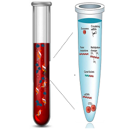
A liquid biopsy is a diagnostic procedure that examines a sample of body fluid, typically blood, to detect cancer. Instead of physically removing tissue from the tumor site (as in a traditional biopsy), liquid biopsies search for circulating tumor DNA (ctDNA), circulating tumor cells (CTCs), and other cancer-related molecules in the bloodstream.

How Liquid Biopsies Work

The basis of liquid biopsies is rooted in the biology of tumors. Cancerous tumors shed cells and DNA fragments into the bloodstream and other body fluids. This circulating tumor DNA (ctDNA) and Circulating Tumor Cells (CTCs) carry genetic mutations that can provide valuable information about the tumor. Liquid biopsies capture these markers and use advanced genomic sequencing technologies to analyze their genetic and molecular properties.

  1. Circulating Tumor DNA (ctDNA): This consists of small fragments of DNA shed into the bloodstream by cancer cells. It carries the genetic mutations of the tumor, enabling an in-depth look at the cancer's genomic profile.
  2. Circulating Tumor Cells (CTCs): CTCs are cancer cells that have detached from the primary tumor and entered the bloodstream. They can lead to the formation of metastatic tumors if they find a suitable environment to grow.

Liquid Biopsies in Solid Tumors

Traditionally, management of solid tumors has been challenging due to difficulties in early detection, tumor heterogeneity, and the dynamic nature of tumors. Here is how liquid biopsies can play a crucial role:

  1. Early Detection: Detecting solid tumors at an early stage improves patient prognosis significantly. Liquid biopsies can identify the presence of cancer-associated mutations in ctDNA or CTCs, potentially even before symptoms or traditional imaging can detect the cancer.
  2. Real-Time Tumor Monitoring: As the cancer progresses or responds to therapy, its genetic makeup can change. This can lead to treatment resistance. Liquid biopsies can track these changes in real-time, offering a more dynamic approach to monitor cancer progression and treatment response.
  3. Therapeutic Guidance: Liquid biopsies can help identify specific mutations driving tumor growth. This information can be used to select targeted therapies and personalize treatment plans. Also, it can help detect acquired resistance to therapies, allowing for timely modifications in the treatment regimen.
  4. Minimal Residual Disease and Recurrence: Liquid biopsies can be used to detect minimal residual disease following cancer treatment, providing a prediction for the likelihood of recurrence. In the event of cancer recurrence, liquid biopsies can help identify the reason for the relapse.

Challenges and Future Directions

Despite the potential of liquid biopsies, challenges remain. Sensitivity and specificity can vary, and the presence of ctDNA or CTCs doesn’t always correlate with the presence of a tumor. False positives and negatives can occur.

Technological advancements and large-scale clinical trials are required to refine these methods and validate their utility. As the technology matures, standardized protocols and clinical guidelines will need to be developed.

Liquid biopsies offer a promising avenue for the management of solid tumors. Their ability to provide real-time, personalized molecular information non-invasively positions them at the forefront of precision oncology. Despite the challenges, with ongoing research and development, they have the potential to revolutionize cancer diagnostics and therapeutics, ushering in a new era in cancer care.

WHO Classification of Brain Tumors and Molecular Changes in Brain Tumors: Emerging Treatment Options for Gliomas


Brain tumors are complex and diverse neoplasms that pose significant challenges in terms of diagnosis and treatment. The World Health Organization (WHO) classification system provides a framework for categorizing brain tumors based on their histopathological features. In recent years, advancements in molecular biology have shed light on the underlying genetic alterations in brain tumors, leading to a better understanding of their biology and paving the way for targeted therapies. This article explores the WHO classification of brain tumors, highlights the molecular changes observed in these tumors, and discusses the emerging treatment options, particularly for gliomas.

WHO Classification of Brain Tumors:

The WHO classification system for brain tumors is a widely accepted and utilized system that provides a standardized approach for classifying these tumors based on their histological characteristics. The most recent edition, the WHO Classification of Tumors of the Central Nervous System 2016, introduced a more integrated approach, incorporating both histopathology and molecular parameters. The classification system stratifies brain tumors into different categories, including gliomas, meningiomas, medulloblastomas, and others, each with its unique subtypes and grades.

Molecular Changes in Brain Tumors:

Advancements in molecular profiling techniques have unraveled the intricate genetic alterations that occur in brain tumors. Gliomas, the most common type of primary brain tumor, have been extensively studied in this regard. The two most prevalent molecular markers in gliomas are IDH (isocitrate dehydrogenase) mutations and 1p/19q co-deletion.

IDH mutations are frequently observed in diffuse gliomas, particularly in lower-grade gliomas (WHO grade II and III). These mutations occur in genes encoding enzymes involved in cellular metabolism, leading to altered metabolic pathways and subsequent tumorigenesis. IDH mutation status has prognostic implications and also guides treatment decisions.

1p/19q co-deletion is a characteristic genetic alteration in oligodendrogliomas, a subtype of gliomas. This molecular abnormality is associated with better response to chemotherapy and improved overall survival. It helps distinguish oligodendrogliomas from other gliomas and influences treatment strategies.

Emerging Treatment Options for Gliomas:

The evolving understanding of molecular changes in gliomas has paved the way for targeted therapies, complementing conventional treatment modalities like surgery, radiation, and chemotherapy. Several promising treatment options are emerging for gliomas, including:

  1. Targeted therapies: Drugs that specifically target molecular alterations in gliomas, such as IDH inhibitors, are being developed and tested in clinical trials. These therapies aim to disrupt the aberrant pathways driving tumor growth while minimizing damage to normal brain tissue.
  2. Immunotherapy: The use of immune checkpoint inhibitors and chimeric antigen receptor (CAR) T-cell therapy has shown promise in the treatment of gliomas. These therapies harness the power of the immune system to recognize and eliminate tumor cells selectively.
  3. Gene therapy: Advances in gene editing technologies, such as CRISPR-Cas9, hold potential for modifying genetic abnormalities in gliomas. Gene therapy approaches are being explored to target and repair specific mutations or inactivate oncogenes to hinder tumor growth.
  4. Personalized medicine: With the advent of molecular profiling, personalized medicine approaches are becoming increasingly relevant. By analyzing the genetic makeup of an individual's tumor, treatment strategies can be tailored to target the specific molecular alterations present, potentially enhancing treatment efficacy.

The WHO classification of brain tumors provides a standardized framework for understanding and categorizing these complex neoplasms. The integration of molecular parameters into the classification system has facilitated a deeper understanding of the underlying genetic alterations in brain tumors. This knowledge has paved the way for the development of targeted therapies and personalized treatment options, particularly for gliomas. As research continues to unravel the intricate molecular changes in brain tumors, further advancements in treatment strategies hold promise for improving outcomes and quality of life for patients with these challenging conditions.

Limitations of Currently Available eGFR Equations (estimated glomerular filtration rate): A Comprehensive Analysis


The estimated glomerular filtration rate (eGFR) is a widely used measure of kidney function that helps assess the filtration capacity of the kidneys. It is an essential parameter for diagnosing and managing various kidney diseases. Several equations have been developed to estimate GFR based on readily available laboratory measurements, such as serum creatinine, age, gender, and race. While these equations have revolutionized the assessment of renal function, it is crucial to recognize their limitations. This article aims to provide a detailed analysis of the limitations associated with currently available eGFR equations.

Population Characteristics:

One of the primary limitations of eGFR equations is their applicability across diverse populations. Many equations were initially developed and validated using predominantly Caucasian populations, which may not accurately reflect GFR in individuals from different ethnic backgrounds. Variations in body composition, muscle mass, dietary habits, and genetic factors can affect serum creatinine levels, leading to inaccurate eGFR estimations.

Age and Gender:

Most eGFR equations incorporate age and gender as variables, assuming a linear relationship between these factors and GFR decline. However, this assumption may not hold true in certain populations. For example, older adults may experience an age-related decline in GFR that is not adequately accounted for by linear equations. Additionally, some equations do not account for gender-specific differences in creatinine metabolism and clearance, potentially leading to inaccuracies in eGFR estimations.

Obesity and Body Composition:

Obesity is a prevalent condition that can significantly impact the accuracy of eGFR equations. Many equations rely on serum creatinine levels, which are influenced by muscle mass. In obese individuals, higher muscle mass can lead to higher creatinine levels, potentially overestimating eGFR. Additionally, variations in body composition, such as increased adipose tissue, may affect the relationship between creatinine production and GFR, further compromising the accuracy of eGFR estimations.

Muscle Wasting and Malnutrition:

Patients with conditions characterized by muscle wasting, such as chronic kidney disease (CKD), liver disease, or cancer, may have reduced muscle mass, resulting in lower creatinine production. Consequently, eGFR equations relying on creatinine levels may underestimate true GFR in these individuals. Malnutrition and low dietary protein intake can also influence serum creatinine levels, leading to inaccurate eGFR estimations.

Kidney Function Variability:

eGFR equations assume a stable relationship between serum creatinine and GFR. However, kidney function can exhibit variability due to factors such as dehydration, medications, or acute illness. Changes in extrarenal creatinine elimination, such as tubular secretion or drug interactions, can affect serum creatinine levels independently of GFR. In such cases, eGFR equations may not accurately reflect true kidney function.

Non-Steady-State Conditions:

eGFR equations are less accurate in non-steady-state conditions, such as acute kidney injury (AKI). Serum creatinine levels may rise rapidly in AKI, while eGFR equations typically estimate GFR based on a steady-state assumption. Consequently, eGFR equations may not provide reliable estimations in patients with fluctuating renal function.

While eGFR equations have undoubtedly improved the assessment of kidney function, it is crucial to recognize their limitations. Population characteristics, age, gender, obesity, body composition, muscle wasting, malnutrition, kidney function variability, and non-steady-state conditions can all contribute to inaccuracies in eGFR estimations. Awareness of these limitations is vital for clinicians to interpret eGFR results appropriately and consider alternative methods, such as direct GFR measurement or adjustment equations, when necessary. 

Importance of Urinalysis in Kidney Diseases


Urinalysis is a key diagnostic tool in the field of nephrology. It involves the examination of urine for various parameters, including color, clarity, concentration, and content (such as glucose, proteins, blood, pH, and various cellular elements). The information obtained from a urinalysis can provide valuable insight into renal function and help identify and monitor kidney diseases.

Importance of Urinalysis in Kidney Diseases:

Detection of Proteinuria: The presence of an abnormal amount of protein in the urine, or proteinuria, is a common indicator of kidney disease. Conditions such as glomerulonephritis, diabetic nephropathy, and nephrotic syndrome can cause significant proteinuria. Urinalysis can quantify protein levels and, along with clinical information, help diagnose these conditions.

Hematuria Identification: Hematuria, the presence of red blood cells in the urine, can be detected through urinalysis. Hematuria can indicate various renal conditions, including urinary tract infections, kidney stones, and more severe disorders like kidney cancers or glomerular diseases.

Identification of Crystals and Casts: The presence of crystals or cellular casts in the urine can suggest specific renal conditions. For instance, red cell casts are indicative of glomerulonephritis, waxy casts suggest advanced kidney disease, and crystals could indicate kidney stones or metabolic disorders.

Glucose and Ketone Measurement: Urinalysis can detect glucose and ketones in the urine. Their presence might indicate poorly controlled diabetes, a condition that can lead to diabetic nephropathy, a leading cause of chronic kidney disease.

Assessment of Kidney Function: Parameters like urine specific gravity and osmolality provide insight into the kidney's concentrating ability, often impaired in chronic kidney diseases.

Clinical Implications of Urinalysis:

Urinalysis serves as an initial, non-invasive screening tool for diagnosing kidney diseases. It is also crucial for monitoring disease progression and response to treatment in conditions like diabetic nephropathy or lupus nephritis. Regular urinalysis can help detect disease flares or relapses, guiding modifications in treatment. Moreover, in the setting of kidney transplantation, urinalysis can help detect early signs of rejection.

Urinalysis plays a vital role in the diagnosis, monitoring, and management of kidney diseases. By providing valuable information about the kidney's functional status and detecting abnormal constituents in urine, it serves as an indispensable tool in nephrology.

What are the expected questions from the above article:

  1. Why is urinalysis an important tool in the diagnosis of kidney diseases?
  2. How can urinalysis help detect proteinuria and what might this indicate about renal health?
  3. What does the presence of hematuria suggest about kidney conditions?
  4. How do crystals and cellular casts in urine contribute to the diagnosis of specific renal disorders?
  5. How can urinalysis be used to monitor the progression of kidney disease and response to treatment?
  6. How does urinalysis contribute to the assessment of kidney function in chronic kidney disease?
  7. What is the role of urinalysis in the context of kidney transplantation?

Pathology of HIV-Associated Nephropathy (HIVAN)


HIV-Associated Nephropathy (HIVAN) is a progressive kidney disease associated with advanced HIV infection. It is one of the most common causes of end-stage renal disease (ESRD) in HIV-infected individuals. The disease is characterized by collapsing focal segmental glomerulosclerosis, tubular dilation, and interstitial inflammation.

Pathology of HIVAN:

HIVAN primarily affects the glomeruli and tubules of the kidneys. The disease is characterized by two distinct pathological changes:

Collapsing Focal Segmental Glomerulosclerosis (FSGS): This is the hallmark of HIVAN, characterized by the collapse and sclerosis of glomerular capillary tufts, along with hyperplasia and hypertrophy of the overlying podocytes. Podocyte injury is a crucial factor in the development of FSGS, and viral proteins from HIV have been shown to directly injure podocytes, leading to proteinuria and progressive renal dysfunction.

Tubulointerstitial disease: This involves tubular dilation, microcyst formation, and interstitial inflammation with infiltration of monocytes and lymphocytes. Tubular epithelial cells also show regenerative changes, with marked hypertrophy, hyperplasia, and mitotic figures. These changes result in progressive renal failure and tubular proteinuria.

HIV infects renal epithelial cells directly, including podocytes and tubular epithelial cells, contributing to the pathogenesis of HIVAN. HIV genes have been found in these cells in individuals with HIVAN, and the expression of HIV proteins in these cells can lead to dysregulation of cell cycle processes, leading to the characteristic pathological changes of the disease.

Clinical Presentation and Management of HIVAN:

HIVAN usually presents in patients with advanced HIV infection or AIDS. The typical clinical features include heavy proteinuria, rapidly progressive renal failure, and large echogenic kidneys on ultrasound. It disproportionately affects individuals of African descent.

The mainstay of treatment for HIVAN is antiretroviral therapy (ART), which can lead to significant improvement in renal function and proteinuria. Other treatment strategies may include angiotensin-converting enzyme (ACE) inhibitors or angiotensin receptor blockers (ARBs) to reduce proteinuria, and dialysis or kidney transplantation for those with ESRD.

HIVAN is a severe complication of HIV infection, leading to significant morbidity and mortality. Understanding the unique pathological changes in the kidneys caused by HIV is critical to the diagnosis and management of this condition. With advances in antiretroviral therapy, the prognosis for patients with HIVAN has improved, but it remains a significant clinical challenge.

What are the expected questions from the above article

  1. What are the characteristic pathological features of HIVAN?
  2. How does HIV infection lead to the development of HIVAN at a cellular level?
  3. What is the role of podocytes and tubular epithelial cells in the pathogenesis of HIVAN?
  4. How does HIVAN typically present clinically?
  5. What are the main treatment strategies for managing HIVAN?
  6. How does antiretroviral therapy influence the course of HIVAN?
  7. What is the impact of HIVAN on the morbidity and mortality of individuals with HIV infection?

Renal Handling of Magnesium: Physiology and Clinical Significance

Magnesium, the second most abundant intracellular cation, plays a vital role in many physiological processes, including energy metabolism, cell growth, and maintaining normal heart rhythm. The kidneys play a critical role in maintaining magnesium homeostasis, which involves processes of filtration, reabsorption, and excretion.

Physiology of Renal Magnesium Handling:

Filtration: Nearly all the magnesium in the plasma is freely filtered at the glomerulus because it exists in an unbound form.

Reabsorption: After filtration, about 95% of magnesium is reabsorbed in the renal tubules, primarily in the thick ascending limb of the loop of Henle (~70%), and to a lesser extent in the distal convoluted tubule (~10-20%) and the proximal tubule (~10-15%). The paracellular pathway is the primary mechanism for magnesium reabsorption in the thick ascending limb, driven by the lumen-positive transepithelial potential difference generated by the active reabsorption of sodium and potassium. In the distal convoluted tubule, magnesium reabsorption is transcellular and regulated by the transient receptor potential melastatin 6 (TRPM6) channel.

Excretion: The remaining magnesium that is not reabsorbed is excreted in urine. The fine-tuning of urinary magnesium excretion occurs mainly in the distal convoluted tubule, and this is influenced by several factors, including plasma magnesium concentration, calcium levels, hormones like aldosterone, and diuretics.

Clinical Significance of Renal Magnesium Handling:

Abnormalities in renal handling of magnesium can lead to magnesium imbalances, which have important clinical implications:

Hypomagnesemia: Reduced renal reabsorption of magnesium can lead to hypomagnesemia (low serum magnesium). This can occur due to genetic defects in magnesium transport (like Gitelman and Bartter syndromes), medications (like diuretics and certain chemotherapeutic drugs), alcoholism, and malnutrition. Symptoms may include neuromuscular irritability, cardiac arrhythmias, and seizures.

Hypermagnesemia: Reduced filtration or increased reabsorption can result in hypermagnesemia (high serum magnesium). This condition is less common and often iatrogenic, related to excessive magnesium intake (like antacids or supplements) in patients with renal insufficiency or failure. Symptoms may include muscle weakness, hypotension, bradycardia, and in severe cases, cardiac arrest.

The kidneys are instrumental in regulating magnesium balance in the body. Understanding the mechanisms of renal magnesium handling and their dysregulation in different pathological states can guide diagnosis, treatment, and management of disorders related to magnesium imbalance.

What are the expected question from the above

  1. How do the kidneys regulate magnesium homeostasis?
  2. Describe the mechanisms of magnesium filtration and reabsorption in the kidneys.
  3. How does the loop of Henle and the distal convoluted tubule contribute to magnesium reabsorption?
  4. What factors influence the fine-tuning of urinary magnesium excretion in the distal convoluted tubule?
  5. Explain the pathophysiological mechanisms that lead to hypomagnesemia and hypermagnesemia.
  6. What are the clinical manifestations of magnesium imbalance and how can they be managed?

Phosphatonins: Physiology and Clinical Significance


Phosphatonins are a group of hormones that play a critical role in phosphate homeostasis, regulating phosphate reabsorption in the renal tubules and contributing to bone mineral metabolism. Their primary function is to inhibit renal phosphate reabsorption, leading to increased phosphate excretion.

Physiology of Phosphatonins:

The most well-known phosphatonin is Fibroblast Growth Factor 23 (FGF23). Produced mainly by osteocytes and osteoblasts in the bone, FGF23 acts on the kidney to reduce phosphate reabsorption and decrease the synthesis of calcitriol (active Vitamin D), which subsequently reduces intestinal phosphate absorption.

FGF23 exerts its effects by binding to the FGF receptor complex in the presence of a co-receptor known as α-Klotho. This interaction activates signaling pathways that lead to decreased expression of the type IIa sodium-phosphate cotransporters (NaPi-IIa) in the proximal renal tubules, resulting in reduced phosphate reabsorption and increased urinary phosphate excretion.

Another key phosphatonin is Secreted Frizzled-Related Protein 4 (sFRP-4). This protein is produced by tumor cells and acts to reduce renal tubular reabsorption of phosphate by downregulating the NaPi-IIa cotransporter, leading to increased phosphate excretion.

Clinical Significance of Phosphatonins:

Alterations in phosphatonin levels can lead to various pathophysiological conditions:

  1. Chronic Kidney Disease (CKD): As kidney function declines in CKD, the ability to excrete phosphate decreases, leading to hyperphosphatemia. To compensate, FGF23 levels rise to decrease renal phosphate reabsorption. Over time, however, persistent high levels of FGF23 can contribute to left ventricular hypertrophy, a significant cause of morbidity and mortality in CKD patients.
  2. Tumor-Induced Osteomalacia (TIO): TIO is a rare paraneoplastic syndrome caused by the overproduction of phosphatonins (mainly FGF23) by tumors. Excess FGF23 leads to hypophosphatemia, reduced calcitriol synthesis, and osteomalacia.
  3. X-linked Hypophosphatemic Rickets (XLH): XLH is a genetic disorder caused by mutations in the PHEX gene, leading to increased FGF23 activity. This causes hypophosphatemia, rickets in children, and osteomalacia in adults.
  4. Autosomal Dominant Hypophosphatemic Rickets (ADHR): ADHR is caused by mutations in the FGF23 gene that make the hormone resistant to degradation. This results in an excess of FGF23, leading to hypophosphatemia, rickets, and osteomalacia.
  5. Phosphatonins play a critical role in phosphate homeostasis and bone health. Understanding their physiology and the pathologies associated with their dysregulation has improved our ability to diagnose and treat disorders of phosphate metabolism. As we continue to explore their mechanisms of action, we may uncover new therapeutic targets for these conditions.

This above answer the below questions:

  1. What are phosphatonins, and what is their primary function in the body?
  2. How does FGF23 regulate phosphate homeostasis?
  3. What role does the co-receptor α-Klotho play in the actions of FGF23?
  4. What is the role of sFRP-4 as a phosphatonin?
  5. How do alterations in phosphatonin levels contribute to the pathophysiology of chronic kidney disease?

Physiology of Solute Removal in Continuous Ambulatory Peritoneal Dialysis

Continuous Ambulatory Peritoneal Dialysis (CAPD) is a type of peritoneal dialysis that allows for the removal of solutes and waste products from the blood when the kidneys are unable to do so. This renal replacement therapy involves the continuous exchange of dialysate within the peritoneal cavity, leveraging the body's natural membranes for filtration.

Physiology of CAPD:

CAPD leverages the patient's peritoneum as a semi-permeable membrane that allows for the exchange of solutes and water. A dialysate solution, rich in glucose, is instilled into the peritoneal cavity. This solution creates an osmotic gradient, facilitating fluid removal, while the peritoneum acts as a membrane allowing solute exchange between blood vessels in the peritoneum and the dialysate.

1. Diffusion: Solute removal in CAPD primarily occurs via diffusion. This is the passive movement of solutes from an area of high concentration to an area of low concentration. In the case of CAPD, toxins such as urea and creatinine in the blood move from the peritoneal capillaries into the dialysate because of the concentration gradient.

2. Ultrafiltration: Fluid removal in CAPD occurs via ultrafiltration. This process is driven by the osmotic gradient created by the high glucose concentration in the dialysate. The high glucose concentration pulls water, along with dissolved solutes, from the blood vessels in the peritoneal cavity into the dialysate.

3. Equilibration: Over time, the concentrations of solutes in the dialysate and the blood equilibrate, meaning they become the same. When this happens, the dialysate is drained and replaced with fresh dialysate, re-establishing the concentration gradients and allowing for further solute removal.

4. Transport Status: Each patient's peritoneum has different permeability characteristics, known as the transport status. High transporters have a high rate of solute and water exchange, while low transporters have a slower rate of exchange. The transport status influences the dialysis prescription, including dwell time (the length of time the dialysate stays in the peritoneal cavity) and the type of dialysate used.

CAPD is a sophisticated process that utilizes the body's natural physiology to clear toxins and excess fluid from the body. Understanding the principles of diffusion, ultrafiltration, and equilibration in the context of an individual's unique peritoneal transport status allows healthcare providers to tailor dialysis treatment to each patient's needs. As we continue to refine our understanding of these processes, we can enhance the efficacy and patient-specific approach of CAPD.

The above will answer the below questions:

  1. Explain how the principles of diffusion and ultrafiltration contribute to solute and fluid removal in CAPD?
  2. What is the role of the peritoneum in CAPD, and how does it function as a semi-permeable membrane?
  3. How does the concentration of glucose in the dialysate facilitate the process of CAPD?
  4. What is equilibration in the context of CAPD, and why does it necessitate the replacement of the dialysate?
  5. How does a patient's transport status influence the CAPD process and the choice of dialysis prescription?
  6. How can understanding the physiology of CAPD inform patient-specific treatment strategies and improve patient outcomes?
  7. What are the potential complications and limitations of CAPD related to the process of solute removal?

The Role of Kidneys in the Pathogenesis of Primary Hypertension


Primary hypertension, also known as essential hypertension, is a multifactorial disease whose exact cause remains largely unknown. However, research has demonstrated that the kidneys play a critical role in the regulation of blood pressure and, therefore, are key players in the pathogenesis of primary hypertension.

Role of Kidneys in Blood Pressure Regulation:

The kidneys participate in blood pressure regulation through several interconnected mechanisms:

1. Sodium Balance: The kidneys control the excretion and reabsorption of sodium. Sodium balance affects the volume of fluid in the blood vessels and, therefore, the blood pressure. A high sodium diet, in some individuals, can lead to increased sodium and fluid retention, resulting in higher blood volume and pressure.

2. Renin-Angiotensin-Aldosterone System (RAAS): The RAAS is a hormonal cascade that plays a key role in blood pressure regulation. In response to decreased blood flow or sodium levels, the kidneys release renin, which triggers a series of reactions leading to the production of angiotensin II and aldosterone. Angiotensin II causes vasoconstriction and promotes the release of aldosterone, which in turn leads to increased sodium and water reabsorption, thereby increasing blood volume and pressure.

3. Pressure-Natriuresis Relationship: This refers to the concept that an increase in arterial pressure leads to an increase in sodium excretion (natriuresis). The ability of the kidneys to excrete excess sodium in response to increases in blood pressure is an important counter-regulatory mechanism. If this mechanism is impaired, as seen in some people with primary hypertension, it can contribute to increased blood pressure.

Kidneys and the Pathogenesis of Primary Hypertension:

Primary hypertension is thought to occur as a result of a complex interplay between genetic, renal, and environmental factors. Here's how the kidneys are involved:

1. Abnormal Sodium Handling: An inability to efficiently excrete dietary sodium is seen in some individuals with primary hypertension. This can result in increased blood volume and blood pressure. While it's not clear why some people have this abnormality, both genetic and environmental factors (such as a high sodium diet) appear to play a role.

2. Altered RAAS Activity: Overactivity of the RAAS can lead to increased vasoconstriction and fluid retention, leading to hypertension. Certain genetic variations can make some individuals more susceptible to this overactivity.

3. Impaired Pressure-Natriuresis: In some individuals with hypertension, the pressure-natriuresis mechanism is shifted to a higher blood pressure. This means that their kidneys do not excrete sodium as efficiently at normal blood pressure levels, leading to increased fluid volume and hypertension.

While the exact pathogenesis of primary hypertension is multifactorial and complex, it is clear that the kidneys play a vital role. They are key regulators of blood pressure and any abnormalities in their function or their response to signals can contribute to the development of hypertension. Understanding the role of the kidneys in hypertension can aid in the development of more targeted treatments for this common condition. Future research may further elucidate these mechanisms and identify novel therapeutic targets for the management of primary hypertension.

The above answered the following 

  1. What is the role of the renin-angiotensin-aldosterone system (RAAS) in blood pressure regulation and how does it contribute to the pathogenesis of primary hypertension?
  2. How does the kidney regulate sodium balance, and how can dysregulation lead to hypertension?
  3. Explain the pressure-natriuresis relationship. How can impairment in this mechanism contribute to the development of hypertension?
  4. What genetic and environmental factors contribute to the pathogenesis of primary hypertension and how do they interact with renal function?
  5. Can you describe some of the current or potential future therapeutic targets for managing primary hypertension that focus on renal mechanisms?