A site for medical students - Practical,Theory,Osce Notes

>
Showing posts with label oncology. Show all posts

Chemical Carcinogens: An In-depth Overview

Chemical carcinogens are substances that can contribute to the development of cancer by causing changes (mutations) in a cell's DNA or leading to other types of cellular damage. These changes can potentially lead to uncontrolled cell division and growth, the hallmarks of cancer. This article provides a comprehensive overview of chemical carcinogens, their sources, mechanisms of action, and strategies to minimize exposure.

Understanding Chemical Carcinogens

Chemical carcinogens are classified based on their ability to initiate or promote cancer. Initiators are chemicals that cause DNA damage and mutations, while promoters stimulate the proliferation of these mutated cells but do not cause mutations themselves. Many chemical carcinogens are capable of both initiating and promoting cancer.

Sources of Chemical Carcinogens

Chemical carcinogens are found in various sources, including:

  1. Tobacco Smoke: This contains over 60 known carcinogens, including polycyclic aromatic hydrocarbons (PAHs), N-nitrosamines, and aldehydes. These are responsible for the strong association between tobacco use and cancers of the lung, mouth, throat, and other organs.
  2. Diet: Certain foods can contain chemical carcinogens. For instance, aflatoxins produced by molds on improperly stored grains and nuts are potent liver carcinogens. Processed meats often contain N-nitroso compounds, while high-temperature cooking can produce heterocyclic amines and PAHs.
  3. Occupational Exposure: Certain industries expose workers to carcinogens, such as asbestos in construction, benzene in chemical manufacturing, and coal tar in metalworking industries.
  4. Environment: Exposure can occur through polluted air, water, or soil. Common environmental carcinogens include asbestos, arsenic, and certain byproducts of industrial processes.

Mechanisms of Action

Chemical carcinogens can damage DNA directly or require metabolic activation to become carcinogenic. For instance, benzene, a known leukemogen, is metabolized in the liver to produce reactive intermediates, which can cause DNA damage leading to leukemia.

Chemical carcinogens can also cause cancer through non-genotoxic mechanisms, such as inducing chronic inflammation, suppressing immune responses, or disrupting cell signaling pathways that regulate cell growth and differentiation.

Mitigating Exposure to Chemical Carcinogens

Reducing exposure to chemical carcinogens is a key strategy in cancer prevention. This can be achieved through various means:

  1. Lifestyle Choices: Avoiding tobacco, limiting alcohol consumption, eating a healthy diet, and exercising regularly can significantly reduce the risk of cancer.
  2. Occupational Safety: Implementing safety regulations and protective measures in workplaces can minimize exposure to occupational carcinogens.
  3. Environmental Regulations: Enforcing laws to control pollution and limit public exposure to environmental carcinogens is crucial.
  4. Education and Awareness: Public awareness campaigns about the risks of carcinogens can encourage healthier behaviors and demand for safer products.

Pain Management in Patients with Spinal Cord Compression


Spinal cord compression (SCC) is a serious condition that occurs when a mass places pressure on the spinal cord. This pressure can originate from various sources, including a herniated disc, a bone fracture, or a tumor. Patients with SCC often experience significant pain, in addition to other neurological symptoms such as weakness or numbness. Consequently, pain management is a critical aspect of care for these patients. This article explores the various strategies and treatments for pain management in patients with spinal cord compression.

Understanding Spinal Cord Compression Pain

The pain associated with SCC can manifest in several ways. It may present as back or neck pain at the site of the compression, radiating pain that spreads to the limbs, or even as a band-like pain around the trunk. This pain can severely affect a patient's quality of life, making effective pain management strategies crucial.

Non-Pharmacological Interventions

Non-drug approaches are often used as adjuncts to medication in managing pain from SCC. These can include:

  1. Physical Therapy: Specific exercises can help alleviate some types of pain and improve mobility and strength.
  2. Occupational Therapy: This can teach coping strategies and modifications to daily activities to help manage pain and improve function.
  3. Psychological Support: Cognitive-behavioral therapy, relaxation techniques, and other psychological interventions can help patients manage the emotional impact of chronic pain.

Pharmacological Management

Medication is often the first line of treatment for SCC-related pain:

  1. Non-Steroidal Anti-Inflammatory Drugs (NSAIDs): These drugs can help reduce pain and inflammation. They are often used for mild to moderate pain.
  2. Corticosteroids: These can reduce inflammation and swelling around the spinal cord, relieving pressure and pain.
  3. Opioids: For severe pain, opioids may be prescribed. However, their use needs careful monitoring due to the risk of dependency and side effects.
  4. Adjuvant Analgesics: Certain antidepressants and anticonvulsants can help manage neuropathic pain often associated with SCC.

Interventional Techniques

When medication and non-drug interventions are insufficient, more invasive strategies may be considered:

  1. Nerve Blocks: These involve injecting medication around specific nerves or into the epidural space to block pain signals.
  2. Neurostimulation Devices: These devices deliver electrical stimulation to the spinal cord or specific nerves to block the perception of pain.
  3. Intrathecal Pumps: These devices deliver pain medication directly to the space around the spinal cord.

Surgical Intervention

In some cases, surgery may be required to remove or reduce the source of the compression. While the primary goal is to alleviate the pressure on the spinal cord, surgery can also significantly reduce pain.

Palliative Care

For patients with advanced disease where the focus is on comfort rather than cure, palliative care plays an essential role. This approach prioritizes quality of life, symptom relief, and psychosocial support.

Pain management in patients with spinal cord compression involves a multifaceted approach, considering non-pharmacological methods, medications, interventional techniques, possible surgical intervention, and palliative care. Ultimately, the goal is to improve the patient's quality of life by effectively managing pain and enhancing overall functionality. Given the complexity of SCC and its associated pain, a personalized, multidisciplinary approach is key to successful management.

Role of Surgery in Abdominal Non-Hodgkin’s Lymphoma


Non-Hodgkin’s lymphoma (NHL) is a heterogeneous group of malignancies of the lymphatic system. While the disease primarily involves lymph nodes, it can also arise in extranodal sites, with the gastrointestinal (GI) tract being the most commonly affected site. Abdominal non-Hodgkin’s lymphoma can involve any part of the GI tract, from the stomach and small intestine to the colon and rectum. The role of surgery in the management of abdominal NHL has evolved significantly over the years and remains a topic of ongoing debate. This article reviews the current understanding and application of surgery in the treatment of abdominal non-Hodgkin’s lymphoma.

Therapeutic Paradigms and the Role of Surgery

Traditionally, the primary modality for treating non-Hodgkin's lymphoma has been chemotherapy, with or without radiotherapy, based on the type and stage of the disease. However, the role of surgery has shifted from a therapeutic to a largely diagnostic and supportive role.

Today, surgical intervention in abdominal NHL is generally reserved for specific situations, including:

  1. Diagnosis and Staging: A biopsy is typically required to confirm the diagnosis of NHL. This may be obtained through endoscopy, image-guided biopsy, or occasionally surgical biopsy if less invasive methods are unsuccessful. Staging laparotomy, once a common practice in lymphoma management, has largely been replaced by less invasive imaging techniques.
  2. Management of Complications: Surgical intervention may be necessary for emergent situations such as bowel perforation, obstruction, hemorrhage, or acute abdomen, which can occur in aggressive cases of abdominal NHL.
  3. Debulking Surgery: The role of debulking surgery (removal of a significant portion of the tumor) is controversial in NHL, as lymphomas are generally considered systemic diseases. However, it may be considered in specific cases, particularly when the disease is localized and causing severe symptoms, or to improve the efficacy of adjuvant therapies.

Risks and Considerations of Surgery

While surgery can offer benefits in certain situations, it's not without potential risks. These can include surgical complications such as infection, bleeding, and damage to nearby organs, as well as longer-term impacts such as bowel dysfunction. Additionally, the recovery time required after surgery can delay the initiation of systemic therapies, which can be detrimental in a disease like NHL that often progresses rapidly.

Therefore, the decision to proceed with surgical intervention should be made carefully, with consideration of the individual patient's disease characteristics, overall health status, and personal wishes. Multidisciplinary discussions involving medical oncologists, radiation oncologists, and surgeons are key to devising an optimal, personalized treatment plan for each patient with abdominal NHL.

The role of surgery in the treatment of abdominal non-Hodgkin’s lymphoma has evolved significantly, with an increased understanding of the disease's systemic nature and advances in systemic therapies. While surgery is no longer a primary treatment modality, it still has an important role in diagnosis, management of complications, and occasionally, debulking. As our knowledge and treatment strategies continue to evolve, the role of surgery will continue to be refined, with the ultimate goal of improving patient outcomes.

Medullary Carcinoma of the Thyroid: A Comprehensive Overview

Medullary thyroid carcinoma (MTC) is a rare but distinctive type of thyroid cancer that originates from parafollicular C cells, which produce the hormone calcitonin. Although it only accounts for about 1-2% of all thyroid cancers, MTC is often aggressive and can pose significant treatment challenges. This article explores the key aspects of MTC, from its pathogenesis to diagnosis and treatment options.

Understanding Medullary Thyroid Carcinoma

MTC is unique among thyroid cancers due to its origin from parafollicular C cells rather than follicular cells, which are the source of the more common papillary and follicular thyroid cancers. Elevated levels of calcitonin, a hormone produced by C cells, serve as a key indicator of MTC.

MTC can occur in two forms: sporadic (non-hereditary) or hereditary. About 75-80% of all MTC cases are sporadic, appearing randomly with no family history. The remaining 20-25% of MTC cases are hereditary and are associated with a genetic syndrome known as Multiple Endocrine Neoplasia type 2 (MEN2).

Signs and Symptoms

The clinical manifestations of MTC can vary. Some patients may notice a lump in their neck, while others may experience symptoms like hoarseness, difficulty swallowing, or a change in voice. Some people with hereditary MTC may not have any symptoms and the disease may be detected through genetic screening.

High levels of calcitonin may lead to symptoms such as diarrhea or flushing. In more advanced cases where the cancer has spread to other parts of the body, symptoms may include bone pain or shortness of breath.

Diagnosis

MTC diagnosis involves a combination of physical examination, imaging studies, laboratory tests, and biopsy.

Elevated calcitonin levels can provide a key indication of MTC. However, a definitive diagnosis requires a biopsy, typically a fine-needle aspiration (FNA), where cells from the thyroid nodule are collected and examined under a microscope.

Imaging tests, such as ultrasound of the neck, computed tomography (CT), or magnetic resonance imaging (MRI), can help define the extent of the disease. For hereditary MTC, genetic testing is done to identify mutations in the RET (rearranged during transfection) proto-oncogene.

Treatment

Surgery is the mainstay of treatment for MTC and often involves a total thyroidectomy, where the entire thyroid gland is removed. In cases where the cancer has spread to nearby lymph nodes, a lymph node dissection may also be performed.

Radioactive iodine, commonly used in other types of thyroid cancer, is generally ineffective in MTC as the C cells do not absorb iodine. Hence, external beam radiation therapy may be considered for certain patients, especially when there is extensive local disease or in cases of recurrence.

For advanced or metastatic disease, systemic therapies may be required. These include chemotherapy, targeted therapies, and immunotherapy. Targeted therapies such as tyrosine kinase inhibitors (like vandetanib and cabozantinib) have shown promise in treating advanced MTC.

Patients with hereditary MTC linked to MEN2 syndrome often undergo prophylactic thyroidectomy, removing the thyroid gland before cancer develops.

Prognosis

The prognosis for MTC varies widely and depends on several factors, including the stage of the disease, the patient's age, and whether the cancer is sporadic or hereditary. Early-stage MTC has a generally good prognosis, with a 10-year survival rate of about 95%. However, for individuals with advanced disease, the prognosis is more guarded, with 10-year survival rates dropping significantly.

Patients with hereditary MTC, especially those diagnosed through genetic screening before the onset of symptoms, often have a better prognosis than those with sporadic MTC. This is largely due to earlier detection and intervention in these cases.

The key to improving prognosis lies in early detection and prompt, appropriate treatment. Therefore, in patients with a known genetic predisposition, regular screening and preventative surgery are important measures.

Medullary thyroid carcinoma, while a small proportion of thyroid cancers, presents unique challenges in diagnosis and treatment due to its origin from parafollicular C cells and its potential to be a hereditary condition.

In the case of hereditary MTC, genetic counseling and testing are paramount in the management of the condition. For both sporadic and hereditary MTC, surgery is the mainstay of treatment, with systemic therapies playing a role in more advanced disease states. While promising strides have been made in understanding and managing MTC, ongoing research is crucial to continue improving outcomes for patients with this unique form of thyroid cancer.

CDK4/6 Inhibitors in Breast Cancer: An In-depth Analysis


Cyclin-dependent kinases 4 and 6 (CDK4/6) inhibitors have revolutionized the treatment landscape for hormone receptor-positive (HR+) metastatic breast cancer. They block proteins that promote cell division and thereby slow cancer growth. This article will delve into the role of CDK4/6 inhibitors in the treatment of breast cancer.

CDK4/6 Inhibition and Its Role in Cell Cycle

Cyclin-dependent kinases 4 and 6 (CDK4/6) are crucial regulators of the cell cycle, which orchestrates cell growth and division. In conjunction with cyclin D, they drive the cell's transition from the G1 phase (the initial growth phase) to the S phase (the DNA synthesis phase). Overactivity of this pathway can lead to unchecked cell proliferation, a hallmark of cancer.

CDK4/6 inhibitors interfere with this process. They bind to CDK4/6 proteins and prevent them from initiating the cell cycle, thereby halting cell division and proliferation. This effect is particularly potent in HR+ breast cancer cells, which are often heavily reliant on the cyclin D-CDK4/6 pathway.

CDK4/6 Inhibitors in Breast Cancer Treatment

Currently, three CDK4/6 inhibitors - palbociclib, ribociclib, and abemaciclib - are approved for use in the treatment of HR+ HER2-negative metastatic breast cancer. These drugs are typically used in combination with endocrine therapy as first or second-line treatment.

  1. Palbociclib (Ibrance): Palbociclib, in combination with letrozole (an aromatase inhibitor), is a standard first-line treatment for postmenopausal women with HR+, HER2- metastatic breast cancer. It can also be used with fulvestrant (a selective estrogen receptor degrader) in women who have progressed after endocrine therapy.
  2. Ribociclib (Kisqali): Ribociclib can be used in combination with an aromatase inhibitor as a first-line treatment for postmenopausal women with HR+, HER2- advanced or metastatic breast cancer. It is also approved for use with fulvestrant in postmenopausal women with HR+, HER2- advanced or metastatic breast cancer as initial endocrine-based therapy or following disease progression on endocrine therapy.
  3. Abemaciclib (Verzenio): Abemaciclib is approved in combination with an aromatase inhibitor as initial endocrine-based therapy for postmenopausal women with HR+, HER2- advanced or metastatic breast cancer. It is also approved for use with fulvestrant in women with disease progression following endocrine therapy.

Efficacy and Safety

Clinical trials have shown that the addition of a CDK4/6 inhibitor to endocrine therapy significantly improves progression-free survival (PFS) in patients with advanced HR+, HER2- breast cancer.

However, like all medicines, CDK4/6 inhibitors can have side effects. Common side effects include neutropenia (low white blood cell count), fatigue, nausea, diarrhea, and alopecia (hair loss). Abemaciclib, unlike the other two inhibitors, commonly causes diarrhea but less neutropenia. Careful patient monitoring and management strategies can mitigate these side effects.


Prophylactic Cranial Irradiation in Small Cell Lung Cancer: A Comprehensive Review


Small Cell Lung Cancer (SCLC) is an aggressive type of lung cancer characterized by rapid growth and a propensity for early metastasis. Despite initial responsiveness to chemotherapy, prognosis remains poor with high rates of relapse. One common site of metastasis is the brain. To combat this, a preventive measure known as Prophylactic Cranial Irradiation (PCI) is often used.

What is Prophylactic Cranial Irradiation (PCI)?

PCI is a preventative treatment strategy in which radiation is administered to the brain to kill potential microscopic cancer cells before they develop into detectable metastatic disease. In SCLC, this is particularly relevant due to the high propensity of this cancer to metastasize to the brain.

Efficacy of PCI in Small Cell Lung Cancer

The utility of PCI in SCLC has been well-documented. A landmark study by the European Organisation for Research and Treatment of Cancer (EORTC) showed that PCI reduced the incidence of symptomatic brain metastases and improved overall survival in patients with SCLC who had responded to initial therapy.

Furthermore, a meta-analysis of individual data from seven randomized clinical trials confirmed a significant reduction in the risk of symptomatic brain metastases and a small but significant improvement in overall survival in patients receiving PCI.

Criteria for Use

PCI is typically considered for patients with SCLC who have responded to initial chemotherapy and radiation therapy, with no evidence of cancer spread to the brain. Before undergoing PCI, patients often undergo brain imaging (MRI or CT) to confirm the absence of brain metastases. However, the use of PCI should be a patient-specific decision that considers the patient’s overall health, performance status, potential side effects, and personal preferences.

Potential Side Effects and Risks

Though PCI can be beneficial, it comes with potential risks and side effects. Common short-term side effects include fatigue, headache, nausea, and hair loss. More concerning are the potential long-term neurocognitive effects. Studies have shown that PCI can lead to memory loss, difficulties in concentration and thinking, and in rare cases, more severe neurological side effects like leukoencephalopathy.

The risk of neurocognitive decline must be weighed against the benefits of PCI in reducing the likelihood of brain metastases. In recent years, there is increasing interest in finding the optimal balance to deliver PCI effectively while minimizing potential neurocognitive impacts.

In summary, PCI remains a key component in the management of SCLC due to its efficacy in reducing the incidence of brain metastases and improving overall survival. However, it is crucial to individualize the decision to administer PCI, considering both the potential benefits and the risk of side effects, including neurocognitive decline. Continued research is needed to optimize the delivery of PCI and mitigate its long-term side effects, ultimately improving the outcomes for patients with SCLC.

Liquid Biopsies in Solid Tumors: A Comprehensive Overview

A paradigm shift in the management and treatment of solid tumors is underway, led by the emergence of 'liquid biopsies.' This non-invasive, revolutionary technology promises to detect cancer, monitor its progress, and guide treatment decisions based on real-time molecular information.

What is a Liquid Biopsy?

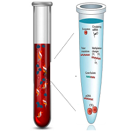
A liquid biopsy is a diagnostic procedure that examines a sample of body fluid, typically blood, to detect cancer. Instead of physically removing tissue from the tumor site (as in a traditional biopsy), liquid biopsies search for circulating tumor DNA (ctDNA), circulating tumor cells (CTCs), and other cancer-related molecules in the bloodstream.

How Liquid Biopsies Work

The basis of liquid biopsies is rooted in the biology of tumors. Cancerous tumors shed cells and DNA fragments into the bloodstream and other body fluids. This circulating tumor DNA (ctDNA) and Circulating Tumor Cells (CTCs) carry genetic mutations that can provide valuable information about the tumor. Liquid biopsies capture these markers and use advanced genomic sequencing technologies to analyze their genetic and molecular properties.

  1. Circulating Tumor DNA (ctDNA): This consists of small fragments of DNA shed into the bloodstream by cancer cells. It carries the genetic mutations of the tumor, enabling an in-depth look at the cancer's genomic profile.
  2. Circulating Tumor Cells (CTCs): CTCs are cancer cells that have detached from the primary tumor and entered the bloodstream. They can lead to the formation of metastatic tumors if they find a suitable environment to grow.

Liquid Biopsies in Solid Tumors

Traditionally, management of solid tumors has been challenging due to difficulties in early detection, tumor heterogeneity, and the dynamic nature of tumors. Here is how liquid biopsies can play a crucial role:

  1. Early Detection: Detecting solid tumors at an early stage improves patient prognosis significantly. Liquid biopsies can identify the presence of cancer-associated mutations in ctDNA or CTCs, potentially even before symptoms or traditional imaging can detect the cancer.
  2. Real-Time Tumor Monitoring: As the cancer progresses or responds to therapy, its genetic makeup can change. This can lead to treatment resistance. Liquid biopsies can track these changes in real-time, offering a more dynamic approach to monitor cancer progression and treatment response.
  3. Therapeutic Guidance: Liquid biopsies can help identify specific mutations driving tumor growth. This information can be used to select targeted therapies and personalize treatment plans. Also, it can help detect acquired resistance to therapies, allowing for timely modifications in the treatment regimen.
  4. Minimal Residual Disease and Recurrence: Liquid biopsies can be used to detect minimal residual disease following cancer treatment, providing a prediction for the likelihood of recurrence. In the event of cancer recurrence, liquid biopsies can help identify the reason for the relapse.

Challenges and Future Directions

Despite the potential of liquid biopsies, challenges remain. Sensitivity and specificity can vary, and the presence of ctDNA or CTCs doesn’t always correlate with the presence of a tumor. False positives and negatives can occur.

Technological advancements and large-scale clinical trials are required to refine these methods and validate their utility. As the technology matures, standardized protocols and clinical guidelines will need to be developed.

Liquid biopsies offer a promising avenue for the management of solid tumors. Their ability to provide real-time, personalized molecular information non-invasively positions them at the forefront of precision oncology. Despite the challenges, with ongoing research and development, they have the potential to revolutionize cancer diagnostics and therapeutics, ushering in a new era in cancer care.

WHO Classification of Brain Tumors and Molecular Changes in Brain Tumors: Emerging Treatment Options for Gliomas


Brain tumors are complex and diverse neoplasms that pose significant challenges in terms of diagnosis and treatment. The World Health Organization (WHO) classification system provides a framework for categorizing brain tumors based on their histopathological features. In recent years, advancements in molecular biology have shed light on the underlying genetic alterations in brain tumors, leading to a better understanding of their biology and paving the way for targeted therapies. This article explores the WHO classification of brain tumors, highlights the molecular changes observed in these tumors, and discusses the emerging treatment options, particularly for gliomas.

WHO Classification of Brain Tumors:

The WHO classification system for brain tumors is a widely accepted and utilized system that provides a standardized approach for classifying these tumors based on their histological characteristics. The most recent edition, the WHO Classification of Tumors of the Central Nervous System 2016, introduced a more integrated approach, incorporating both histopathology and molecular parameters. The classification system stratifies brain tumors into different categories, including gliomas, meningiomas, medulloblastomas, and others, each with its unique subtypes and grades.

Molecular Changes in Brain Tumors:

Advancements in molecular profiling techniques have unraveled the intricate genetic alterations that occur in brain tumors. Gliomas, the most common type of primary brain tumor, have been extensively studied in this regard. The two most prevalent molecular markers in gliomas are IDH (isocitrate dehydrogenase) mutations and 1p/19q co-deletion.

IDH mutations are frequently observed in diffuse gliomas, particularly in lower-grade gliomas (WHO grade II and III). These mutations occur in genes encoding enzymes involved in cellular metabolism, leading to altered metabolic pathways and subsequent tumorigenesis. IDH mutation status has prognostic implications and also guides treatment decisions.

1p/19q co-deletion is a characteristic genetic alteration in oligodendrogliomas, a subtype of gliomas. This molecular abnormality is associated with better response to chemotherapy and improved overall survival. It helps distinguish oligodendrogliomas from other gliomas and influences treatment strategies.

Emerging Treatment Options for Gliomas:

The evolving understanding of molecular changes in gliomas has paved the way for targeted therapies, complementing conventional treatment modalities like surgery, radiation, and chemotherapy. Several promising treatment options are emerging for gliomas, including:

  1. Targeted therapies: Drugs that specifically target molecular alterations in gliomas, such as IDH inhibitors, are being developed and tested in clinical trials. These therapies aim to disrupt the aberrant pathways driving tumor growth while minimizing damage to normal brain tissue.
  2. Immunotherapy: The use of immune checkpoint inhibitors and chimeric antigen receptor (CAR) T-cell therapy has shown promise in the treatment of gliomas. These therapies harness the power of the immune system to recognize and eliminate tumor cells selectively.
  3. Gene therapy: Advances in gene editing technologies, such as CRISPR-Cas9, hold potential for modifying genetic abnormalities in gliomas. Gene therapy approaches are being explored to target and repair specific mutations or inactivate oncogenes to hinder tumor growth.
  4. Personalized medicine: With the advent of molecular profiling, personalized medicine approaches are becoming increasingly relevant. By analyzing the genetic makeup of an individual's tumor, treatment strategies can be tailored to target the specific molecular alterations present, potentially enhancing treatment efficacy.

The WHO classification of brain tumors provides a standardized framework for understanding and categorizing these complex neoplasms. The integration of molecular parameters into the classification system has facilitated a deeper understanding of the underlying genetic alterations in brain tumors. This knowledge has paved the way for the development of targeted therapies and personalized treatment options, particularly for gliomas. As research continues to unravel the intricate molecular changes in brain tumors, further advancements in treatment strategies hold promise for improving outcomes and quality of life for patients with these challenging conditions.