Non-compaction of the left ventricle (NCLV), also known as left ventricular non-compaction (LVNC), is a rare congenital cardiac abnormality characterized by prominent trabeculations and deep recesses in the myocardium of the left ventricle (LV). In this response, we will discuss the embryology, anatomy, and physiology of NCLV in detail.
Embryology:
During embryonic development, the heart undergoes complex morphological changes. Initially, the heart tube forms, and the cardiac myocytes begin to differentiate. Subsequently, the heart undergoes looping, leading to the formation of the four-chambered heart. During the looping process, trabeculations develop within the myocardium. Over time, these trabeculations normally become compacted, resulting in a smooth endocardial surface.
Anatomy:
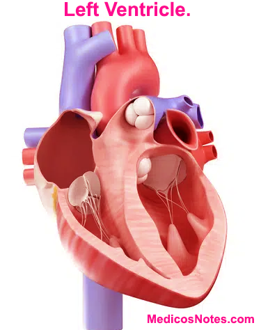
In NCLV, the normal compaction of the myocardium does not occur, leading to the persistence of excessive trabeculations and deep recesses. The non-compacted myocardium is characterized by multiple, prominent trabeculations separated by deep intertrabecular recesses. These recesses communicate with the LV cavity, creating a sponge-like appearance. The non-compacted region is typically located in the mid-lateral or apical segments of the LV, but it can involve other areas as well.
Physiology:
The physiology of NCLV is not fully understood, but it is believed to result from impaired myocardial development during embryogenesis. The non-compacted myocardium is thought to be functionally compromised due to several factors:
Impaired contractility: The trabeculations within the non-compacted myocardium are not efficiently contractile, leading to reduced systolic function. This results in a decreased ejection fraction and impaired ventricular emptying.
Ventricular stiffness: The deep intertrabecular recesses can restrict blood flow within the non-compacted myocardium, leading to increased ventricular stiffness. This stiffness further impairs ventricular filling during diastole, reducing the overall cardiac output.
Electrophysiological abnormalities: NCLV is often associated with arrhythmias and conduction abnormalities. The distorted architecture of the myocardium disrupts the normal electrical conduction pathways, leading to arrhythmias such as ventricular tachycardia, ventricular fibrillation, and atrioventricular block.
Embolic events: The recesses and trabeculations within the LV can act as stagnant blood pools, predisposing individuals with NCLV to thrombus formation. Thrombi can embolize to systemic circulation, leading to stroke or other systemic embolic events.
It is important to note that NCLV can be associated with other structural heart defects, such as mitral valve abnormalities, ventricular septal defects, and coarctation of the aorta. These additional abnormalities further contribute to the clinical manifestations and severity of the condition.
In summary, non-compaction of the left ventricle is a congenital cardiac abnormality characterized by excessive trabeculations and deep recesses within the myocardium. The impaired compaction during embryogenesis leads to functional abnormalities, including impaired contractility, ventricular stiffness, electrophysiological abnormalities, and increased risk of embolic events. Understanding the embryology, anatomy, and physiology of NCLV is crucial for the diagnosis, management, and appropriate treatment of this rare condition.


