A site for medical students - Practical,Theory,Osce Notes

>
Showing posts with label neurology. Show all posts

Hirayama Disease - Investigation and MRI findings


Investigating Hirayama disease involves various diagnostic tests to confirm the clinical suspicion and differentiate it from other neuromuscular disorders. Magnetic resonance imaging (MRI) plays a crucial role in the evaluation of Hirayama disease, providing specific findings that are characteristic of this condition. Let's explore the investigations and MRI findings associated with Hirayama disease:

Clinical Evaluation: The initial step in diagnosing Hirayama disease involves a thorough clinical assessment, focusing on the characteristic clinical features such as muscle weakness and atrophy, asymmetry of involvement, cold paresis, and preservation of sensory functions.

Electromyography (EMG) and Nerve Conduction Studies (NCS): EMG and NCS may be performed to evaluate the electrical activity of muscles and nerve conduction in the affected limbs. These tests can help exclude other neuromuscular disorders and confirm the diagnosis of a lower motor neuron pathology.

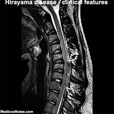
Magnetic Resonance Imaging (MRI): MRI is a key investigation in the diagnosis of Hirayama disease. The imaging findings are typically observed in the cervical spinal cord region, specifically the lower cervical segments (C5-C7). The following MRI findings are characteristic of Hirayama disease:

a. Forward Displacement of the Dural Sac: MRI often reveals a forward displacement of the dural sac during neck flexion. This occurs due to dynamic compression and elongation of the lower cervical spinal cord.

b. Posterior Epidural Space Enlargement: Enlargement of the posterior epidural space, located behind the dural sac, is a common finding in Hirayama disease. This can be visualized as a crescent-shaped area of CSF (cerebrospinal fluid) accumulation.

c. Flattening of the Spinal Cord: MRI may show flattening or concavity of the lower cervical spinal cord, particularly on the posterior aspect, during neck flexion. This is caused by compression of the spinal cord against the anterior vertebral column.

d. Vertebral Column Abnormalities: In some cases, MRI may reveal mild structural abnormalities in the vertebral column, such as focal kyphosis or loss of normal cervical lordosis. These abnormalities contribute to the spinal cord compression during neck flexion.

e. Normal Signal Intensity: Despite the clinical weakness and atrophy observed in Hirayama disease, MRI typically shows normal signal intensity of the affected spinal cord segments. This finding distinguishes it from other progressive motor neuron disorders characterized by abnormal signal intensity on MRI.

It is important to note that the MRI findings may be more pronounced during active neck flexion and may appear normal or less significant during neutral or extended neck positions. Thus, performing MRI with neck flexion is crucial to detect the characteristic findings associated with Hirayama disease.

While clinical evaluation and MRI findings are key in the diagnosis of Hirayama disease, additional investigations, such as laboratory tests and genetic studies, may be performed to exclude other conditions and assess for underlying genetic predispositions. The combination of clinical assessment and MRI findings is essential for a definitive diagnosis of Hirayama disease, enabling appropriate management and support for affected individuals.

Hirayama disease - Treatment

Hirayama Disease - Investigation and MRI findings
Hirayama disease - Clinical Features - An Overview

Understanding Hirayama Disease, Ethiopathogenesis

Hirayama disease - Clinical Features - An Overview

Hirayama disease, also known as juvenile nonprogressive cervical amyotrophy or monomelic amyotrophy, is a rare neurological disorder characterized by distinct clinical features. These features are essential for diagnosing and differentiating Hirayama disease from other neuromuscular conditions. 

Explore the clinical features commonly associated with Hirayama disease:

Age of Onset: Hirayama disease typically manifests in young individuals, predominantly males, in their second or third decade of life. The age of onset ranges from 10 to 25 years, with a peak incidence during adolescence.

Muscle Weakness and Atrophy: The hallmark symptom of Hirayama disease is muscle weakness and atrophy, primarily affecting the muscles of the hand and forearm on one side of the body. The weakness usually begins insidiously and progresses slowly over weeks to months. The atrophy may be more noticeable in the muscles on the back of the hand, leading to a "claw-like" appearance.

Asymmetric Involvement: Hirayama disease typically exhibits asymmetric involvement, with one side of the body being more affected than the other. The weakness and atrophy usually start unilaterally and may eventually spread to involve the other side, although to a lesser extent. This asymmetry helps distinguish Hirayama disease from other progressive neuromuscular disorders.

Muscle Cramps and Fasciculations: Some individuals with Hirayama disease may experience muscle cramps, especially during activity. Fasciculations, which are involuntary muscle twitches or contractions, can also be observed in the affected muscles.

Cold Paresis: A characteristic feature of Hirayama disease is the phenomenon of "cold paresis." Patients may notice worsening of muscle weakness and coordination issues in cold environments or during exposure to cold objects. This symptom is believed to be related to the altered blood flow dynamics and ischemia in the affected spinal cord region.

Sensory Function Preservation: Sensory functions are typically spared in Hirayama disease. Patients do not experience significant sensory loss or abnormalities in the affected limbs. This is another important feature that helps distinguish it from other disorders involving both motor and sensory deficits.

Nonprogressive Course: Hirayama disease is known for its nonprogressive nature. Once the symptoms stabilize, which usually occurs within a few years of onset, further progression is rare. However, it is important to note that the initial progression phase may last several months to a few years before stabilization.

Normal Deep Tendon Reflexes: Deep tendon reflexes, such as the biceps and triceps reflexes, are typically preserved or only mildly affected in Hirayama disease. This finding distinguishes it from other motor neuron disorders characterized by hyperactive or absent reflexes.

It is important to consider these clinical features along with additional diagnostic tests, such as electromyography (EMG), nerve conduction studies, and magnetic resonance imaging (MRI), to arrive at an accurate diagnosis of Hirayama disease. Early recognition and appropriate management can help improve the quality of life for individuals affected by this rare neurological disorder.

Hirayama disease - Treatment

Hirayama Disease - Investigation and MRI findings

Hirayama disease - Clinical Features - An Overview

Understanding Hirayama Disease, Ethiopathogenesis

Understanding Hirayama Disease, ethiopathogenesis


Hirayama disease, also known as juvenile nonprogressive cervical amyotrophy or monomelic amyotrophy, is a rare neurological disorder characterized by muscle weakness and wasting, primarily affecting the muscles of the hand and forearm. The ethiopathogenesis, or the causes and mechanisms, of Hirayama disease have been the subject of scientific investigation, although the exact etiology remains partially understood.

Understanding Hirayama Disease:

Hirayama disease typically affects young individuals, predominantly males, in their second or third decade of life. The condition is more commonly observed in Asian populations, particularly in Japan and India. The clinical course of Hirayama disease is usually nonprogressive, meaning the symptoms do not worsen over time.

Ethiopathogenesis of Hirayama Disease:

Dynamic Compression of the Lower Cervical Spinal Cord: One of the primary theories regarding the ethiopathogenesis of Hirayama disease is dynamic compression of the lower cervical spinal cord. During neck flexion, there is a shift of the posterior dural sac, resulting in anterior displacement and compression of the spinal cord. This compression leads to ischemia and damage to the anterior horns of the spinal cord, causing muscle weakness and atrophy.

Circulatory Disturbances and Vascular Factors: Vascular factors have also been implicated in the development of Hirayama disease. It is believed that the abnormal posture adopted by individuals with this condition, which involves neck flexion and the chin tucking inwards, leads to mechanical compression of the vertebral arteries and subsequent disturbances in blood flow. This compromised blood supply can result in ischemia and neuronal damage in the lower cervical spinal cord.

Genetic Predisposition: Genetic factors may contribute to the development of Hirayama disease. Certain genetic variations and human leukocyte antigen (HLA) associations have been identified in affected individuals, suggesting a potential genetic predisposition. For instance, the HLA-DRB11501 and HLA-DRB11502 haplotypes have been found to be more prevalent in Hirayama disease patients. These genetic factors may influence the susceptibility to spinal cord compression and vascular disturbances.

Immunological Factors: Some studies have proposed an immunological component in the ethiopathogenesis of Hirayama disease. Autoantibodies against various neuronal antigens, such as heat shock proteins, have been detected in the serum and cerebrospinal fluid of affected individuals. These autoantibodies may trigger an autoimmune response that contributes to the damage observed in the lower cervical spinal cord.

Hirayama disease is a rare neurological disorder characterized by muscle weakness and wasting, primarily affecting the muscles of the hand and forearm. The ethiopathogenesis of this condition involves dynamic compression of the lower cervical spinal cord, circulatory disturbances and vascular factors, genetic predisposition, and potential immunological factors. While significant progress has been made in understanding the underlying causes and mechanisms of Hirayama disease, further research is necessary to unravel the complex interplay of these factors and develop more targeted diagnostic and therapeutic strategies for affected individuals.

Hirayama disease - Treatment

Hirayama Disease - Investigation and MRI findings

Hirayama disease - Clinical Features - An Overview

Understanding Hirayama Disease, Ethiopathogenesis


Synaptic vesicles 2 (SV2) - An Overview


Synaptic vesicles 2 (SV2) is a protein that is found in synaptic vesicles, which are small membrane-bound organelles that store and release neurotransmitters at the synapse, the junction between two neurons. There are three isoforms of SV2: SV2A, SV2B, and SV2C. SV2A is the most widely expressed isoform and is present in most central nervous system synapses.

Structure: SV2 is a transmembrane glycoprotein that spans the synaptic vesicle membrane. It is composed of 12 transmembrane domains, with its N- and C-termini located on the cytosolic side of the membrane. The glycosylation of SV2 is thought to be important for its function in regulating neurotransmitter release.

Function: SV2 plays a critical role in regulating neurotransmitter release from synaptic vesicles. It is thought to function as a transporter, helping to load neurotransmitters into synaptic vesicles, and as a modulator of the release process. Specifically, SV2 is thought to regulate the size and frequency of neurotransmitter release events, which are important for normal neurotransmission and synaptic plasticity.

Neurological application: Because of its critical role in regulating neurotransmitter release, SV2 has been the target of drugs used in the treatment of neurological disorders such as epilepsy. Specifically, levetiracetam, a widely used antiepileptic drug, is thought to bind to SV2A and modulate its function, leading to a reduction in neurotransmitter release and a decrease in seizure activity. Other drugs that target SV2A are currently being developed for the treatment of other neurological disorders, such as Parkinson's disease and Alzheimer's disease.

In summary, Synaptic Vesicles 2 (SV2) is a protein that plays a critical role in regulating neurotransmitter release from synaptic vesicles. It is a transmembrane glycoprotein that helps to load neurotransmitters into synaptic vesicles and modulate the release process. Because of its role in regulating neurotransmitter release, SV2 has been the target of drugs used in the treatment of neurological disorders such as epilepsy, Parkinson's disease, and Alzheimer's disease.

Structure Function, type and Neurological Disease associated with sodium channels


Sodium channels are specialized proteins that are found on the membranes of many types of cells in the body, including neurons. They play a critical role in generating and propagating electrical signals that are important for normal nervous system function.

Structure: Sodium channels are composed of a large protein molecule that spans the cell membrane, with a pore that allows sodium ions to pass through. The pore is lined with amino acid residues that are critical for regulating the flow of sodium ions. Sodium channels are made up of multiple subunits, with the α subunit forming the central pore and the other subunits contributing to the overall structure and function of the channel.

Function: Sodium channels are responsible for the influx of sodium ions into cells in response to a stimulus, such as a depolarizing electrical signal. This influx of sodium ions triggers the depolarization of the cell, which can in turn lead to the generation of an action potential, or electrical signal, that can be propagated along the axon of a neuron.

Types: There are several different types of sodium channels that are expressed in different tissues and have distinct properties. The most well-known types of sodium channels are Nav1.1 to Nav1.9, which are expressed in different types of neurons and have varying properties. For example, Nav1.7 is important for pain sensation and is a target for analgesic drugs, while Nav1.5 is important for normal heart function and is a target for antiarrhythmic drugs.

Neurological diseases associated with sodium channels: Mutations in genes that encode for sodium channels can lead to a number of neurological diseases. For example, mutations in the SCN1A gene, which encodes for the Nav1.1 sodium channel, can lead to a range of epileptic disorders, including Dravet syndrome, a severe form of childhood epilepsy. Mutations in other sodium channel genes, such as SCN2A, SCN8A, and SCN9A, can also lead to different types of epilepsy, as well as other neurological disorders such as autism spectrum disorder and developmental delay.

Other neurological diseases associated with sodium channels include periodic paralysis, a condition in which affected individuals experience episodes of muscle weakness or paralysis, and myotonia, a condition in which the muscles are slow to relax after contracting. These conditions are caused by mutations in genes that encode for sodium channels expressed in muscle cells.

In summary, sodium channels play a critical role in generating and propagating electrical signals in neurons and other types of cells. There are several different types of sodium channels with distinct properties and functions, and mutations in genes that encode for sodium channels can lead to a range of neurological diseases, including epilepsy, periodic paralysis, and myotonia.

Neurological disease associated Polyglucosan body : Structure, formation and pathophysiology


Polyglucosan bodies, also known as Lafora bodies, are abnormal structures that form in the cytoplasm of cells, particularly in neurons and muscle cells. These structures are composed of abnormal accumulations of a complex carbohydrate called polyglucosan.

Formation: Polyglucosan bodies are formed due to mutations in genes that regulate glycogen metabolism. Normally, glycogen is a branched chain of glucose molecules that serves as a source of energy for the body. However, in individuals with certain genetic mutations, the glycogen molecules are not properly broken down and can accumulate to form polyglucosan bodies.

Pathophysiology: The accumulation of polyglucosan bodies in neurons and muscle cells can lead to a number of problems. In neurons, polyglucosan bodies can disrupt normal cellular functions, leading to cell death and neurological dysfunction. In muscle cells, the accumulation of polyglucosan bodies can interfere with normal muscle contraction and lead to muscle weakness and atrophy.

Neurological disease: One of the most well-known neurological diseases associated with polyglucosan bodies is Lafora disease. Lafora disease is a rare, inherited form of progressive myoclonic epilepsy that typically begins in adolescence. It is caused by mutations in genes that regulate glycogen metabolism, leading to the formation of polyglucosan bodies in the brain. The accumulation of these structures can lead to a wide range of neurological symptoms, including seizures, myoclonus (sudden muscle jerks), and cognitive decline.

Other neurological diseases associated with polyglucosan bodies include adult polyglucosan body disease and glycogen storage disease type IV. These conditions can also lead to neurological symptoms such as muscle weakness, ataxia (loss of coordination), and neuropathy (nerve damage).

In summary, the accumulation of polyglucosan bodies in neurons and muscle cells can lead to a number of problems, particularly neurological dysfunction. Lafora disease is the most well-known neurological disease associated with polyglucosan bodies, but other conditions can also lead to similar symptoms.

Neural synaptic proteins, its function and role in neurological disease


Neural synaptic proteins are a class of proteins that are located at the synapses, which are the sites of communication between neurons. These proteins play a critical role in regulating the transmission of signals across the synapse and are essential for normal neuronal function.

Function: There are several different types of neural synaptic proteins, each with a specific function. Some examples of these proteins include:

1. Neurotransmitter receptors: These proteins are located on the postsynaptic membrane and are responsible for binding neurotransmitters released by the presynaptic neuron. Once a neurotransmitter binds to its receptor, it can either excite or inhibit the postsynaptic neuron, depending on the type of receptor.

2. Synaptic vesicle proteins: These proteins are located in the presynaptic neuron and are responsible for packaging neurotransmitters into vesicles and releasing them into the synapse upon neuronal activation.

3. Synaptic scaffolding proteins: These proteins are located on both the pre- and postsynaptic membranes and are involved in organizing the synaptic structure and regulating the function of other synaptic proteins.

Role in neurological disease: Disruptions in the function of neural synaptic proteins have been implicated in several neurological diseases, including Alzheimer's disease, Parkinson's disease, and schizophrenia. For example, abnormalities in the function of neurotransmitter receptors have been associated with Alzheimer's disease and schizophrenia, while changes in the levels of synaptic scaffolding proteins have been observed in Parkinson's disease. In addition, mutations in genes that encode synaptic proteins have been linked to several neurodevelopmental disorders, such as autism spectrum disorder and intellectual disability. Understanding the role of neural synaptic proteins in neurological diseases may lead to the development of new treatments that target these proteins to alleviate symptoms and improve quality of life for affected individuals.

Structure ,formation, genetics pathophysiology and disease associated with lafora bodies

Lafora bodies are abnormal structures that are found in the neurons and other cells of individuals with Lafora disease. Lafora disease is a rare, inherited form of epilepsy that usually begins in adolescence and leads to progressive neurological deterioration.

Structure: Lafora bodies are abnormal accumulations of glycogen, a complex sugar that is normally stored in cells and used as a source of energy. In individuals with Lafora disease, glycogen accumulates in the form of insoluble, protein-bound inclusions that are known as Lafora bodies. Lafora bodies are irregularly shaped, membrane-bound structures that are composed of abnormal glycogen and other proteins.

Formation: The formation of Lafora bodies is thought to be due to mutations in two genes, EPM2A and NHLRC1, which encode proteins that are involved in glycogen metabolism. Mutations in these genes lead to abnormal glycogen accumulation and the formation of Lafora bodies in cells. The exact mechanism by which Lafora bodies form is not fully understood, but it is thought to involve abnormal interactions between glycogen and other proteins.

Genetics: Lafora disease is an autosomal recessive disorder, meaning that it occurs when an individual inherits two mutated copies of either the EPM2A or NHLRC1 gene, one from each parent. The mutations in these genes lead to abnormal glycogen accumulation and the formation of Lafora bodies in cells.

Pathophysiology: The accumulation of Lafora bodies in cells leads to impaired cellular function and cell death, particularly in neurons in the brain. The exact mechanisms by which Lafora bodies lead to neurological symptoms are not fully understood, but it is thought to involve disruption of cellular signaling and metabolism.

Disease associated with Lafora bodies: Lafora disease is a rare, inherited form of epilepsy that usually begins in adolescence and leads to progressive neurological deterioration. The symptoms of Lafora disease can include seizures, myoclonus (involuntary muscle twitches), cognitive decline, and dementia. There is currently no cure for Lafora disease, and treatment is primarily focused on symptom management.

Structure function ,type and neurological disease associated with potassium channels

Potassium channels are a class of ion channels that play an important role in regulating the electrical activity of cells, including neurons. They allow the selective flow of potassium ions across the cell membrane, which helps to establish and maintain the resting membrane potential of cells.

Structure: Potassium channels are transmembrane proteins that consist of four subunits, each containing six transmembrane helices. The subunits come together to form a pore that allows the selective passage of potassium ions across the cell membrane. There are different types of potassium channels, which differ in their structure, regulation, and function.

Function: Potassium channels help to regulate the electrical activity of cells by controlling the flow of potassium ions across the cell membrane. They play a critical role in shaping the action potential of neurons, which is the electrical signal that allows neurons to communicate with one another. By regulating the flow of potassium ions, potassium channels help to control the excitability of neurons and the frequency and timing of action potentials.

Types of potassium channels: There are several types of potassium channels, including voltage-gated potassium channels, ligand-gated potassium channels, and inward-rectifying potassium channels. Voltage-gated potassium channels open in response to changes in membrane potential, while ligand-gated potassium channels are activated by the binding of specific ligands, such as neurotransmitters. Inward-rectifying potassium channels are important for setting the resting membrane potential of cells.

Neurological diseases associated with potassium channels: Disruptions in potassium channel function have been associated with several neurological diseases, including epilepsy, ataxia, and channelopathies. In channelopathies, genetic mutations in potassium channel genes can lead to abnormal potassium channel function, which can result in neurological symptoms such as muscle weakness, paralysis, and seizures. In some cases, drugs that target specific potassium channels have been developed as treatments for neurological diseases, such as certain forms of epilepsy.


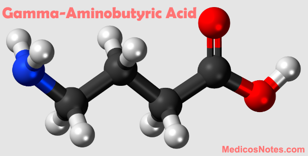
. GABA (gamma-aminobutyric acid) structure ,synthesis ,function and neurological disease associated


GABA, or gamma-aminobutyric acid, is a neurotransmitter in the central nervous system that is involved in regulating neuronal activity. It is an inhibitory neurotransmitter, meaning that it reduces the activity of the neurons it acts upon.

Structure: GABA is an amino acid with a chemical structure similar to glutamate, another neurotransmitter in the brain. It consists of a carboxylic acid group, an amino group, and a side chain that contains a four-carbon chain terminated with an amino group.

Synthesis: GABA is synthesized from glutamate through the action of the enzyme glutamic acid decarboxylase (GAD), which removes a carboxyl group from glutamate. This process requires vitamin B6 as a cofactor. Once synthesized, GABA is packaged into synaptic vesicles and released into the synaptic cleft upon neuronal activation.

Function: GABA plays an important role in regulating neuronal activity and maintaining the balance between excitation and inhibition in the brain. When GABA binds to its receptors on the postsynaptic neuron, it opens chloride ion channels, which leads to hyperpolarization of the cell membrane and a decrease in the likelihood of the neuron firing an action potential.

Neurological diseases associated with GABA: Disruptions in GABAergic neurotransmission have been implicated in several neurological and psychiatric disorders, including anxiety, depression, epilepsy, and schizophrenia. For example, decreased GABA levels or function have been associated with anxiety disorders, while increased GABA levels have been observed in epilepsy. Altered GABAergic function has also been implicated in the development of drug addiction and withdrawal.

Enumerate the excitatory neurotrasmitters and their role in various neurological disease

Excitatory neurotransmitters are chemicals that transmit signals between neurons in the brain and other parts of the nervous system, and they play important roles in various neurological diseases. Here are some examples of excitatory neurotransmitters and their roles in neurological disease:

1. Glutamate: Glutamate is the primary excitatory neurotransmitter in the brain, and it plays an important role in various neurological diseases, such as stroke, epilepsy, and neurodegenerative diseases like Alzheimer's and Parkinson's disease. In these diseases, the release of excess glutamate can lead to excessive activation of excitatory receptors, resulting in neuronal damage and cell death.

2. Acetylcholine: Acetylcholine is an excitatory neurotransmitter that is involved in cognitive processes such as learning and memory, and it is also involved in motor function. In diseases like Alzheimer's disease, there is a loss of acetylcholine neurons in the brain, leading to a reduction in cognitive and motor function.

3. Dopamine: Dopamine is an excitatory neurotransmitter that is involved in reward, motivation, and movement. In diseases like Parkinson's disease, there is a loss of dopamine neurons in the brain, leading to motor symptoms such as tremors, rigidity, and bradykinesia.

4. Norepinephrine: Norepinephrine is an excitatory neurotransmitter that is involved in the stress response, arousal, and attention. In diseases like depression, there is a decrease in norepinephrine levels, leading to a reduction in motivation, energy, and attention.

5. Histamine: Histamine is an excitatory neurotransmitter that is involved in the regulation of sleep, wakefulness, and appetite. In diseases like narcolepsy, there is a dysfunction in histamine neurons, leading to excessive daytime sleepiness and disrupted sleep-wake cycles.

Histamine: The Multifaceted Biogenic Amine in Immune Response, Inflammation, and Neurotransmission

Histamine, a biogenic amine, plays critical roles in immune response, inflammation, and neurotransmission. It is involved in various physiological processes, including the regulation of gastric acid secretion, vasodilation, and bronchoconstriction. This article delves into the synthesis, metabolism, functions, receptor subtypes, and involvement of histamine in physiological processes and various disorders.

1. Synthesis and Metabolism of Histamine

Histamine is synthesized through a single-step process involving the precursor amino acid histidine:

1.1. Synthesis

Histidine decarboxylase (HDC) catalyzes the conversion of histidine to histamine.

1.2. Metabolism

Histamine is metabolized primarily by two enzymes:

Histamine N-methyltransferase (HNMT) converts histamine to N-methylhistamine.

Diamine oxidase (DAO) oxidizes histamine to form imidazole acetaldehyde.

2. Histamine Receptors and Signaling

Histamine receptors are G-protein-coupled receptors (GPCRs) that are classified into four main subtypes:

2.1. H1 Receptors

H1 receptors are involved in smooth muscle contraction, vasodilation, and increased vascular permeability. They are primarily responsible for the symptoms of allergic reactions.

2.2. H2 Receptors

H2 receptors regulate gastric acid secretion, smooth muscle relaxation, and have a role in immune response modulation.

2.3. H3 Receptors

H3 receptors function as autoreceptors and heteroreceptors, regulating the release of histamine and other neurotransmitters in the central nervous system (CNS).

2.4. H4 Receptors

H4 receptors are primarily expressed on immune cells and modulate immune response and inflammation.

3. Major Roles of Histamine

Histamine plays several essential roles in the body, including:

3.1. Immune Response and Inflammation

Histamine is released by mast cells and basophils during an immune response, contributing to inflammation and allergic reactions.

3.2. Gastric Acid Secretion

Histamine stimulates gastric acid secretion by parietal cells in the stomach through the activation of H2 receptors.

3.3. Neurotransmission

Histamine functions as a neurotransmitter in the CNS, modulating arousal, wakefulness, and cognition.

4. Histamine's Involvement in Allergic Reactions

Histamine is a critical mediator of allergic reactions, responsible for symptoms such as itching, redness, swelling, and bronchoconstriction. It is released from mast cells and basophils upon exposure to allergens, leading to the activation of H1 receptors and the initiation of an inflammatory response.

5. Histamine Dysregulation and Associated Disorders

Imbalances in histamine signaling are implicated in various disorders, including:

5.1. Allergic Rhinitis and Asthma

Histamine is a key player in the pathophysiology of allergic rhinitis and asthma, contributing to inflammation and bronchoconstriction.

5.2. Urticaria and Atopic Dermatitis

Elevated histamine levels contribute to the symptoms of urticaria (hives) and atopic dermatitis (eczema), such as itching and inflammation.

5.3. Gastroesophageal Reflux Disease (GERD) and Peptic Ulcers

Overproduction of gastric acid, mediated by histamine's action on H2 receptors, can contribute to the development of GERD and peptic ulcers.

5.4. Histamine Intolerance

Histamine intolerance results from an imbalance between histamine intake and the body's ability to metabolize it. This can lead to a wide range of symptoms, including headaches, gastrointestinal issues, and skin rashes.

6. Therapeutic Approaches Targeting Histamine

Several therapeutic strategies have been developed to modulate histamine signaling, including:

6.1. H1 Receptor Antagonists (Antihistamines)
Antihistamines, such as diphenhydramine, cetirizine, and fexofenadine, block H1 receptors and are commonly used to treat allergic reactions, including allergic rhinitis, urticaria, and atopic dermatitis.

6.2. H2 Receptor Antagonists
H2 receptor antagonists, such as ranitidine, famotidine, and cimetidine, inhibit gastric acid secretion and are used to treat GERD and peptic ulcers.

6.3. Mast Cell Stabilizers
Mast cell stabilizers, such as cromolyn sodium, prevent the release of histamine from mast cells, reducing inflammation and symptoms in conditions like asthma and allergic rhinitis.

Norepinephrine: The Versatile Neurotransmitter Regulating Attention, Arousal, and Stress Response

Norepinephrine (NE), also known as noradrenaline, is a vital neurotransmitter in both the central and peripheral nervous systems. It plays essential roles in attention, arousal, stress response, and cardiovascular regulation. This article delves into the synthesis, metabolism, functions, receptor subtypes, and involvement of norepinephrine in physiological processes and various disorders.

1. Synthesis and Metabolism of Norepinephrine

Norepinephrine is synthesized through a multi-step process involving the precursor amino acid tyrosine:

1.1. Synthesis

Tyrosine hydroxylase (TH) catalyzes the conversion of tyrosine to L-DOPA (L-3,4-dihydroxyphenylalanine).

Aromatic L-amino acid decarboxylase (AADC) converts L-DOPA to dopamine.

Dopamine β-hydroxylase (DBH) converts dopamine to norepinephrine.

1.2. Metabolism

Norepinephrine is metabolized primarily by two enzymes:

Monoamine oxidase (MAO) breaks down norepinephrine into 3,4-dihydroxymandelic acid (DOMA).

Catechol-O-methyltransferase (COMT) converts norepinephrine to normetanephrine.

Norepinephrine Receptors and Signaling

2. Norepinephrine receptors are G-protein-coupled receptors (GPCRs) that are classified into two main families:

2.1. Alpha-Adrenergic Receptors (α1 and α2)

These receptors consist of three α1 subtypes (α1A, α1B, and α1D) and three α2 subtypes (α2A, α2B, and α2C). They mediate various effects on smooth muscle contraction, neurotransmitter release, and other cellular functions.

2.2. Beta-Adrenergic Receptors (β1, β2, and β3)

These receptors stimulate adenylyl cyclase, increasing intracellular cyclic adenosine monophosphate (cAMP) levels and activating protein kinase A (PKA). They regulate processes such as cardiac function, smooth muscle relaxation, and metabolic pathways.

3. Major Noradrenergic Pathways

The primary noradrenergic pathways in the brain include:

3.1. Locus Coeruleus (LC) Pathway

The locus coeruleus is the primary source of norepinephrine in the brain. It projects to various regions, including the cortex, hippocampus, amygdala, and thalamus, modulating attention, arousal, and stress response.

4. Norepinephrine's Role in Attention and Arousal

Norepinephrine plays a crucial role in regulating attention, arousal, and vigilance. It modulates the signal-to-noise ratio of neuronal activity, enhancing the processing of relevant information and suppressing irrelevant information.

5. Norepinephrine's Role in Stress Response

Norepinephrine is a critical component of the body's stress response. It increases heart rate, blood pressure, and blood flow to muscles, preparing the body for a "fight or flight" response. In the brain, it modulates the stress-induced activation of the hypothalamic-pituitary-adrenal (HPA) axis and promotes adaptive behavioral responses.

6. Norepinephrine Dysregulation and Associated Disorders

Imbalances in norepinephrine signaling are implicated in various neurological and psychiatric disorders, including:

6.1. Attention Deficit Hyperactivity Disorder (ADHD)

ADHD has been linked to dysregulation of norepinephrine signaling, particularly in the prefrontal cortex. Medications such as atomoxetine, a selective norepinephrine reuptake inhibitor, are used to treat ADHD by increasing norepinephrine levels.

6.2. Depression

Depression is associated with altered norepinephrine transmission. Some antidepressant medications, such as norepinephrine reuptake inhibitors (NRIs) and norepinephrine-dopamine reuptake inhibitors (NDRIs), act by increasing norepinephrine levels in the synaptic cleft.

6.3. Anxiety Disorders

Excessive noradrenergic activity has been implicated in the pathophysiology of anxiety disorders. Some anxiolytic medications, such as beta-blockers, target norepinephrine receptors to alleviate symptoms of anxiety.

6.4. Post-Traumatic Stress Disorder (PTSD)

Increased norepinephrine signaling has been implicated in the development of PTSD. Some medications, like prazosin, an α1-adrenergic receptor antagonist, are used to treat PTSD-related nightmares and sleep disturbances.

7. Therapeutic Approaches Targeting Norepinephrine

Several therapeutic strategies have been developed to modulate norepinephrine signaling, including:

7.1. Norepinephrine Reuptake Inhibitors (NRIs)

NRIs, such as atomoxetine, block the norepinephrine transporter (NET) and increase norepinephrine levels in the synaptic cleft. They are used to treat ADHD and some forms of depression.

7.2. Alpha-Adrenergic Receptor Antagonists

Drugs like prazosin, which target α1-adrenergic receptors, can be used to treat PTSD-related symptoms, hypertension, and benign prostatic hyperplasia.

7.3. Beta-Adrenergic Receptor Antagonists (Beta-Blockers)

Beta-blockers, such as propranolol and metoprolol, target β-adrenergic receptors and are used to treat hypertension, anxiety, and other cardiovascular disorders.

Dopamine: The Multifunctional Neurotransmitter Shaping Motivation, Reward, and Movement

Dopamine, an essential neurotransmitter in the central nervous system (CNS), plays a vital role in various processes, including motivation, reward, movement, and cognition. This article provides an in-depth exploration of dopamine's synthesis, metabolism, functions, receptor subtypes, and its involvement in both physiological processes and neurological disorders.

1. Synthesis and Metabolism of Dopamine

Dopamine is synthesized through a multi-step process involving the precursor amino acid tyrosine:

1.1. Synthesis

Tyrosine hydroxylase (TH) catalyzes the conversion of tyrosine to L-DOPA (L-3,4-dihydroxyphenylalanine).

Aromatic L-amino acid decarboxylase (AADC) converts L-DOPA to dopamine.

1.2. Metabolism

Dopamine is metabolized mainly by two enzymes:

  1. Monoamine oxidase (MAO) breaks down dopamine into 3,4-dihydroxyphenylacetaldehyde (DOPAL).
  2. Catechol-O-methyltransferase (COMT) converts dopamine to 3-methoxytyramine.
  3. Dopamine Receptors and Signaling

2. Dopamine receptors are G-protein-coupled receptors (GPCRs) that are classified into two main families:

2.1. D1-like Receptors (D1 and D5)

These receptors stimulate adenylyl cyclase, increasing intracellular cyclic adenosine monophosphate (cAMP) levels, and activating protein kinase A (PKA).

2.2. D2-like Receptors (D2, D3, and D4)

These receptors inhibit adenylyl cyclase, reducing cAMP levels and decreasing PKA activity. D2 receptors also function as autoreceptors, regulating dopamine release and synthesis.

3. Major Dopaminergic Pathways

There are several major dopaminergic pathways in the brain:

3.1. Nigrostriatal Pathway

This pathway originates in the substantia nigra pars compacta (SNc) and projects to the striatum. It is primarily involved in the regulation of motor function.

3.2. Mesolimbic Pathway

Originating in the ventral tegmental area (VTA) and projecting to the nucleus accumbens (NAc), this pathway is associated with reward, motivation, and addiction.

3.3. Mesocortical Pathway

This pathway also originates in the VTA but projects to the prefrontal cortex. It is involved in cognitive functions, including attention, working memory, and decision-making.

4. Dopamine's Role in Movement

Dopamine is crucial for the initiation and control of voluntary movement. Imbalances in dopamine signaling in the basal ganglia contribute to movement disorders such as Parkinson's disease.

5. Dopamine's Role in Motivation and Reward

Dopamine plays a central role in the brain's reward system, regulating motivation, reinforcement, and the experience of pleasure. It contributes to the encoding of reward prediction errors, helping to guide decision-making and goal-directed behavior.

6. Dopamine's Role in Cognition

Dopamine modulates cognitive processes, such as attention, working memory, and executive function, primarily through its actions in the prefrontal cortex.

7. Dopamine Dysregulation and Associated Disorders

Imbalances in dopamine signaling are implicated in various neurological and psychiatric disorders, including:

7.1. Parkinson's Disease

Dopamine depletion in the nigrostriatal pathway due to the degeneration of dopeneration of dopaminergic neurons in the substantia nigra pars compacta is a key feature of Parkinson's disease. This results in motor symptoms such as tremors, rigidity, bradykinesia, and postural instability. Dopamine replacement therapies, such as L-DOPA, are the primary treatment for Parkinson's disease.

7.2. Schizophrenia

Dopaminergic dysfunction, particularly hyperactivity in the mesolimbic pathway and hypoactivity in the mesocortical pathway, has been implicated in the development of schizophrenia. Antipsychotic drugs primarily target dopamine D2 receptors to alleviate positive symptoms of schizophrenia, such as hallucinations and delusions.

7.3. Attention Deficit Hyperactivity Disorder (ADHD)

ADHD has been linked to dysregulation of dopamine signaling, particularly in the prefrontal cortex. Stimulant medications like methylphenidate and amphetamine, which increase dopamine levels, are commonly used to treat ADHD.

7.4. Drug Addiction

Drugs of abuse, such as cocaine, amphetamines, and opioids, lead to increased dopamine release in the nucleus accumbens, reinforcing drug-seeking behavior and contributing to addiction.

8. Therapeutic Approaches Targeting Dopamine

Given dopamine's involvement in various neurological and psychiatric disorders, several therapeutic approaches have been developed to target dopamine signaling:

8.1. Dopamine Precursors

L-DOPA, a precursor of dopamine, is used as the gold standard treatment for Parkinson's disease, as it can cross the blood-brain barrier and be converted to dopamine in the brain.

8.2. Dopamine Receptor Agonists and Antagonists

Compounds that selectively target dopamine receptor subtypes can be used to treat various disorders. For example, D2 receptor antagonists are used to treat schizophrenia, while D1/D5 receptor agonists are being investigated for their potential to enhance cognitive function.

8.3. Dopamine Reuptake Inhibitors

Drugs that inhibit the dopamine transporter (DAT) and block dopamine reuptake, such as methylphenidate, are used in the treatment of ADHD and narcolepsy.

8.4. Monoamine Oxidase Inhibitors (MAOIs)

MAOIs, which block the breakdown of dopamine, can be used to treat depression and Parkinson's disease.

Dopamine is a crucial neurotransmitter involved in diverse physiological processes in the brain, including movement, motivation, reward, and cognition. Dysregulation of dopamine signaling is implicated in various neurological and psychiatric disorders, making it an attractive target for therapeutic interventions. As our understanding of dopamine's diverse roles in the CNS deepens, the development of novel and targeted treatments for these disorders becomes increasingly promising.

Acetylcholine: The Multifaceted Neurotransmitter in Learning, Memory, and Muscle Control

Acetylcholine (ACh) is a critical neurotransmitter that plays crucial roles in cognitive function, muscle control, and the regulation of the autonomic nervous system. This article will explore the synthesis, metabolism, and functions of acetylcholine, its receptor subtypes, and its involvement in various physiological processes and disorders.

1. Synthesis and Metabolism of Acetylcholine

Acetylcholine is synthesized in neurons through a single-step process:

1.1. Synthesis

The enzyme choline acetyltransferase (ChAT) catalyzes the transfer of an acetyl group from acetyl-CoA to choline, producing acetylcholine. Choline is transported into neurons via high-affinity choline transporters (CHTs) located on the cell membrane.

1.2. Metabolism

Acetylcholinesterase (AChE) is the primary enzyme responsible for the breakdown of acetylcholine into choline and acetate. This rapid hydrolysis occurs in the synaptic cleft, ensuring that acetylcholine's action is brief and confined.

2. Acetylcholine Receptors and Signaling

Acetylcholine receptors are proteins that bind to acetylcholine and mediate its effects. They are divided into two major classes:

2.1. Nicotinic Acetylcholine Receptors (nAChRs)

These ligand-gated ion channels are activated by acetylcholine binding, allowing cations (mainly Na+ and K+) to pass through the membrane. nAChRs are found in the neuromuscular junction, autonomic ganglia, and the central nervous system. They are also the target of nicotine, hence their name.

2.2. Muscarinic Acetylcholine Receptors (mAChRs)

These G-protein-coupled receptors (GPCRs) modulate ion channels and intracellular signaling pathways indirectly through second messengers. Five subtypes of mAChRs (M1-M5) have been identified, which are widely expressed in the central and peripheral nervous systems.

3. Functions of Acetylcholine in the Central Nervous System

Acetylcholine plays diverse roles in the central nervous system, including:

3.1. Learning and Memory

ACh is essential for cognitive processes such as learning and memory, particularly in the hippocampus and cortex. It contributes to synaptic plasticity, which underlies these processes.

3.2. Attention and Alertness

ACh release in the cortex and thalamus modulates attention and alertness, contributing to the regulation of sleep-wake cycles and the encoding of sensory information.

4. Functions of Acetylcholine in the Peripheral Nervous System

In the peripheral nervous system, acetylcholine has crucial roles:

4.1. Neuromuscular Transmission

ACh is the primary neurotransmitter at the neuromuscular junction, where it activates nAChRs on muscle cells, leading to muscle contraction.

4.2. Autonomic Nervous System

ACh is the primary neurotransmitter in both the sympathetic and parasympathetic branches of the autonomic nervous system. It mediates various physiological responses, such as heart rate regulation, digestion, and pupillary constriction.

5. Acetylcholine Dysregulation and Associated Disorders

Imbalances in acetylcholine signaling have been implicated in various disorders, including:

5.1. Alzheimer's Disease

ACh deficiency, particularly in the cortex and hippocampus, is a hallmark of Alzheimer's disease. Cholinesterase inhibitors, which prevent ACh breakdown , are used as a treatment strategy to enhance cholinergic transmission and alleviate cognitive symptoms in patients with Alzheimer's disease.

5.2. Myasthenia Gravis

Myasthenia gravis is an autoimmune disorder characterized by muscle weakness and fatigue. It results from the production of antibodies that target nAChRs at the neuromuscular junction, impairing neuromuscular transmission. Treatments for myasthenia gravis include cholinesterase inhibitors and immunosuppressive therapies.

5.3. Parkinson's Disease

Although primarily associated with dopamine deficiency, cholinergic dysfunction also contributes to the cognitive and motor symptoms of Parkinson's disease. Anticholinergic drugs are sometimes used to alleviate motor symptoms in patients with Parkinson's disease.

5.4. Schizophrenia

Altered cholinergic signaling, particularly involving the M1 and M4 mAChR subtypes, has been implicated in schizophrenia. Targeting these receptor subtypes may represent a novel therapeutic approach for this psychiatric disorder.

6. Therapeutic Approaches Targeting Acetylcholine

Various therapeutic strategies have been developed to modulate acetylcholine signaling, including:

6.1. Cholinesterase Inhibitors

Drugs like donepezil, rivastigmine, and galantamine inhibit AChE, increasing ACh levels in the synaptic cleft. These medications are primarily used to treat Alzheimer's disease and, in some cases, myasthenia gravis.

6.2. Nicotinic Receptor Agonists and Antagonists

Compounds that target specific nAChR subtypes have potential therapeutic applications for conditions like nicotine addiction, cognitive impairment, and pain management.

6.3. Muscarinic Receptor Modulators

Selective agonists and antagonists for mAChR subtypes are being investigated for their potential use in treating disorders like schizophrenia, Alzheimer's disease, and Parkinson's disease.

Acetylcholine is a versatile neurotransmitter involved in a wide array of physiological processes in both the central and peripheral nervous systems. Its roles in learning, memory, attention, neuromuscular transmission, and autonomic regulation highlight its importance in maintaining proper brain and body function. Dysregulation of acetylcholine signaling is implicated in various neurological and psychiatric disorders, making it a valuable target for therapeutic interventions. As our understanding of acetylcholine's diverse functions continues to grow, so does the potential for the development of novel and targeted treatments for these disorders.

Glutamate: The Essential Neurotransmitter for Learning and Memory

Glutamate, a powerful excitatory neurotransmitter, is indispensable for the proper functioning of the brain. It is involved in a wide range of processes, such as learning, memory, cognition, and neuroplasticity. This article delves into the structure, function, and roles of glutamate in the nervous system, as well as its potential therapeutic applications.

1. The Structure and Synthesis of Glutamate

Glutamate is an amino acid that serves as the most abundant neurotransmitter in the central nervous system (CNS). It has a structure similar to that of other amino acids, consisting of an alpha carbon bonded to an amino group, a carboxyl group, and a unique side chain. In the case of glutamate, the side chain contains an additional carboxyl group, making it an acidic amino acid.

The synthesis of glutamate occurs through two primary pathways:

1.1. Glutaminase Pathway

The enzyme glutaminase catalyzes the conversion of glutamine to glutamate. This pathway is the primary source of glutamate in neurons.

1.2. Transamination Pathway

This pathway involves the enzyme aspartate aminotransferase, which transfers an amino group from aspartate to α-ketoglutarate, producing glutamate and oxaloacetate. This pathway is prominent in astrocytes, which play a crucial role in glutamate homeostasis in the brain.

2. Glutamate Receptors and Signaling

Glutamate receptors are proteins that bind to glutamate and are essential for signal transmission. They are classified into two major groups:

2.1. Ionotropic Glutamate Receptors

These receptors are ligand-gated ion channels that open upon binding to glutamate, allowing ions to flow through the membrane. There are three main subtypes: N-methyl-D-aspartate (NMDA), α-amino-3-hydroxy-5-methyl-4-isoxazolepropionic acid (AMPA), and kainate receptors.

2.2. Metabotropic Glutamate Receptors

These G-protein coupled receptors (GPCRs) modulate ion channels indirectly through intracellular signaling cascades. There are eight subtypes (mGluR1-mGluR8) divided into three groups based on sequence homology and signaling pathways.

3. The Role of Glutamate in Learning and Memory

Glutamate is critical for long-term potentiation (LTP), a process that strengthens synaptic connections in response to repeated stimulation. LTP is thought to underlie learning and memory. The NMDA receptor plays a pivotal role in LTP induction by allowing calcium influx when activated by glutamate and postsynaptic depolarization. This influx triggers intracellular signaling cascades, ultimately resulting in the strengthening of synaptic connections.

4. Glutamate in Neuroplasticity and Neurodevelopment

Glutamate contributes to neuroplasticity, the brain's ability to adapt and reorganize in response to experience. This process is crucial during development, as glutamate signaling guides the formation and refinement of neuronal connections. Glutamate also modulates adult neurogenesis, the birth of new neurons in specific regions of the adult brain, which is implicated in learning and memory.

5. Glutamate Dysregulation and Neurological Disorders

Imbalances in glutamate signaling can contribute to various neurological disorders, including:

5.1. Alzheimer's Disease

Glutamate excitotoxicity, where excessive glutamate overstimulates and damages neurons, is implicated in the progression of Alzheimer's disease. Some treatments aim to modulate glutamate signaling to protect neurons and slow disease progression.

5.2. Parkinson's Disease

Glutamate dysfunction is also implicated in the loss of dopaminergic neurons in the substantia nigra, which characterizes Parkinson's disease. Modulating glutamate transmission may be a promising therapeutic strategy for this disorder.

5.3. Amyotrophic Lateral Sclerosis (ALS)

ALS, a progressive neurodegenerative disease affecting motor neurons, has been linked to glutamate excitotoxicity. Riluzole, an FDA-approved medication for ALS, is thought to act by reducing glutamate release.

5.4. Epilepsy

Excessive glutamate activity can lead to seizures, a hallmark of epilepsy. Some antiepileptic drugs aim to reduce glutamate signaling to control seizures.

5.5. Schizophrenia

Imbalances in glutamate signaling, particularly at the level of NMDA receptors, are implicated in schizophrenia. Novel therapeutic approaches targeting glutamate receptors are being investigated to treat this psychiatric disorder.

5.6. Major Depressive Disorder

Altered glutamate signaling is implicated in the pathophysiology of major depressive disorder. The discovery of rapid-acting antidepressant effects of the NMDA receptor antagonist ketamine has led to a growing interest in glutamatergic-based therapies for depression.

6. Therapeutic Approaches Targeting Glutamate

Given glutamate's involvement in various neurological disorders, several therapeutic approaches have been developed to target glutamate signaling:

6.1. NMDA Receptor Antagonists

Compounds like ketamine and memantine can block NMDA receptors and have demonstrated effectiveness in treating depression and Alzheimer's disease, respectively.

6.2. AMPA Receptor Modulators

Positive allosteric modulators of AMPA receptors, such as aniracetam and CX-516, have shown promise in enhancing cognitive function and treating neurological disorders.

6.3. Metabotropic Glutamate Receptor Modulators

Ligands that target specific metabotropic glutamate receptor subtypes have potential therapeutic applications in treating disorders like Parkinson's disease, anxiety, and depression.

Glutamate is a vital neurotransmitter involved in numerous physiological processes in the brain, including learning, memory, and neuroplasticity. Dysregulation of glutamate signaling is implicated in various neurological and psychiatric disorders, making it an attractive target for therapeutic interventions. As our understanding of glutamate's diverse roles in the CNS deepens, the development of novel and targeted treatments for these disorders becomes increasingly promising.

The ANO3 Gene: Function, Implications, and Research Advances

The human genome is composed of thousands of genes, each with its specific function in the body. Among these genes is the ANO3 gene, which encodes for the Anoctamin 3 protein, also known as Transmembrane Protein 16C (TMEM16C). ANO3 is a member of the Anoctamin family of proteins, which play important roles in regulating ion channels and membrane transport. Recent studies have identified ANO3 gene variations associated with several neurological disorders, including dystonia, Tourette syndrome, and schizophrenia. In this article, we will explore the function of the ANO3 gene, its implications in health and disease, and the latest research advancements in the field.

Function of the ANO3 Gene:

The ANO3 gene is located on chromosome 11 and is composed of 23 exons, which code for a 901 amino acid protein. Anoctamin 3 is a transmembrane protein that is mainly expressed in the brain, particularly in the basal ganglia, a group of structures involved in motor control. The protein is also expressed in other parts of the body, including the pancreas, kidneys, and heart.

Anoctamin 3 has been shown to act as a calcium-activated chloride channel, meaning that it can regulate the flow of chloride ions across the cell membrane in response to changes in intracellular calcium levels. This function is essential for various physiological processes, including muscle contraction, secretion, and neurotransmission. Anoctamin 3 has also been implicated in the regulation of the excitability of neurons in the basal ganglia, which is crucial for controlling movement.

Implications of ANO3 Gene Variations:

Several studies have identified ANO3 gene variations associated with various neurological disorders. For instance, a rare missense variant in ANO3 has been linked to autosomal dominant craniocervical dystonia, a movement disorder characterized by abnormal contractions of the neck and face muscles. Another ANO3 variant has been associated with Tourette syndrome, a neurodevelopmental disorder characterized by repetitive involuntary movements and vocalizations.

Recent studies have also identified ANO3 gene variants associated with schizophrenia, a severe mental illness characterized by disturbances in perception, thought, and behavior. One study found that ANO3 gene variants were more common in individuals with schizophrenia than in controls. Another study found that ANO3 expression was decreased in the prefrontal cortex of individuals with schizophrenia.

Research Advances:

The identification of ANO3 gene variations associated with various neurological disorders has spurred research aimed at understanding the function of the gene and the mechanisms by which ANO3 variants contribute to disease. Recent studies have used animal models and cell cultures to investigate the role of ANO3 in regulating neuronal excitability and movement control.

For instance, a study in mice found that Anoctamin 3 is involved in regulating the activity of a specific type of neuron in the basal ganglia that is essential for controlling movement. Another study in human cells found that ANO3 variants associated with craniocervical dystonia and Tourette syndrome altered the protein's ability to regulate calcium-activated chloride channels.

The ANO3 gene encodes for a transmembrane protein that plays important roles in regulating ion channels and membrane transport. ANO3 gene variations have been associated with several neurological disorders, including dystonia, Tourette syndrome, and schizophrenia. The identification of ANO3 gene variants has spurred research aimed at understanding the function of the gene and the mechanisms by which ANO3 variants contribute to disease. 

VPS13A Gene: Structure, Function, and Clinical Significance

The VPS13A gene encodes the Vacuolar Protein Sorting 13 Homolog A, a protein involved in intracellular trafficking and mitochondrial membrane maintenance. Mutations in the VPS13A gene have been linked to the rare neurodegenerative disorder, Chorea-Acanthocytosis (ChAc). This article will provide a comprehensive overview of the VPS13A gene, its role in cellular function, and the clinical implications of its mutations.

VPS13A gene structure and location

The VPS13A gene is located on the long arm of chromosome 9, specifically at position 9q21.2. It spans over 240 kilobases and consists of 73 exons. The gene is highly conserved among species, signifying its importance in cellular functions.

VPS13A protein function

The VPS13A protein is a member of the VPS13 family, which is involved in various aspects of intracellular trafficking and membrane dynamics. The precise function of VPS13A remains unclear, but studies have suggested that it plays a crucial role in maintaining mitochondrial membrane integrity and lipid homeostasis.

VPS13A is primarily localized to the endoplasmic reticulum (ER) and the outer mitochondrial membrane, where it forms contact sites between these two organelles. This interaction is essential for the transfer of lipids, particularly phospholipids, between the ER and mitochondria. In addition, VPS13A has been implicated in the regulation of autophagy, a process that removes damaged cellular components, including damaged mitochondria, through lysosomal degradation.

Chorea-Acanthocytosis (ChAc) and VPS13A gene mutations

Chorea-Acanthocytosis (ChAc) is a rare, autosomal recessive neurodegenerative disorder characterized by involuntary movements (chorea), abnormal red blood cell morphology (acanthocytosis), and progressive neurological decline. Mutations in the VPS13A gene are responsible for ChAc, with over 70 different pathogenic mutations identified to date. These mutations result in the loss of VPS13A protein function and typically lead to premature truncation or degradation of the protein.

Pathogenesis of ChAc

Although the precise pathogenic mechanisms underlying ChAc remain poorly understood, several hypotheses have been proposed:

a. Mitochondrial dysfunction: Loss of VPS13A function disrupts the lipid homeostasis between the ER and mitochondria, leading to impaired mitochondrial function and increased oxidative stress. This may contribute to neuronal vulnerability and degeneration.

b. Impaired autophagy: VPS13A mutations may disrupt the regulation of autophagy, leading to the accumulation of damaged cellular components and promoting neuronal cell death.

c. Cytoskeletal abnormalities: Acanthocytosis, a key feature of ChAc, results from alterations in the red blood cell membrane and cytoskeleton. It is hypothesized that similar cytoskeletal abnormalities may occur in neurons, contributing to their dysfunction and degeneration.

Diagnosis and management of ChAc

The diagnosis of ChAc relies on the identification of characteristic clinical features, detection of acanthocytosis in peripheral blood smears, and confirmation of VPS13A gene mutations through genetic testing. Currently, there is no cure for ChAc, and management is focused on symptomatic treatment and supportive care. This includes pharmacological interventions to manage chorea and psychiatric symptoms, as well as physical, occupational, and speech therapy to address functional impairments.

Chromosome, Gene, Number of repeats in CTG and significance

CTG repeats refer to a specific type of repetitive DNA sequence where the nucleotides cytosine (C), thymine (T), and guanine (G) repeat in a specific order. The length of these repeats can vary among individuals and is typically measured in terms of the number of CTG repeats.

Chromosome:

The location of the CTG repeat can vary depending on the disorder that it is associated with. For example, in myotonic dystrophy type 1 (DM1), the CTG repeat expansion is located in the 3' untranslated region of the DMPK gene on chromosome 19.

Gene:

The CTG repeat expansion is typically located within the coding or non-coding region of a specific gene. For example, in DM1, the CTG repeat expansion is located in the 3' untranslated region of the DMPK gene, which leads to abnormal RNA splicing and a toxic gain-of-function of the DMPK protein.

Number of repeats:

The number of CTG repeats can vary widely among individuals. In the case of DM1, the normal range of CTG repeats is 5-37. However, individuals with DM1 have CTG repeats that are expanded beyond the normal range, with the size of the expansion correlating with disease severity. For example, individuals with DM1 with fewer than 50 CTG repeats typically have a milder form of the disorder, while those with more than 1,000 CTG repeats have a severe form.

Significance:

The significance of CTG repeat expansions can vary depending on the location of the repeat and the gene that it affects. In the case of DM1, the CTG repeat expansion leads to the production of an abnormal DMPK protein that accumulates in muscle and other tissues, leading to muscle weakness, myotonia (delayed muscle relaxation), and other symptoms. CTG repeat expansions have also been associated with other disorders, such as Huntington's disease, spinocerebellar ataxia, and various forms of muscular dystrophy.

In addition to causing disease, CTG repeat expansions can also have functional consequences. For example, a recent study has shown that CTG repeat expansions in the intron of the TCF4 gene can regulate the expression of nearby genes, leading to altered gene expression and cellular function. Overall, CTG repeat expansions are an important area of research in the study of genetic disorders and gene regulation.

Chromosome, Gene, Protein, Pathogenesis of and epilepsy and epilepsy syndrome

Epilepsy is a neurological disorder characterized by recurrent seizures, which are caused by abnormal electrical activity in the brain. There are many different types of epilepsy, and the genetics of the disorder can be complex.

Chromosome:  

There are several chromosomes that have been implicated in the development of epilepsy. For example, mutations in the SCN1A gene, located on chromosome 2, have been associated with Dravet syndrome, a severe form of epilepsy that typically begins in infancy. Mutations in the DEPDC5 gene, located on chromosome 22, have been associated with familial focal epilepsy with variable foci, a form of epilepsy that tends to run in families.

Gene: 

There are many genes that have been associated with the development of epilepsy. In addition to SCN1A and DEPDC5, mutations in the KCNQ2, KCNQ3, and SCN2A genes have all been associated with different forms of epilepsy. These genes are involved in the regulation of ion channels in the brain, which are critical for the normal functioning of neurons.

Protein: 

The proteins that are involved in the pathogenesis of epilepsy are varied and complex. For example, the sodium channel protein Nav1.1, encoded by the SCN1A gene, is critical for the normal function of inhibitory interneurons in the brain, and mutations in this protein can lead to hyperexcitability of neurons and seizures. The potassium channel proteins KCNQ2 and KCNQ3 are also involved in the regulation of neuronal excitability, and mutations in these proteins can lead to epilepsy.

Pathogenesis: 

The pathogenesis of epilepsy is complex and can be caused by a variety of factors. In some cases, epilepsy is caused by structural abnormalities in the brain, such as tumors or malformations. In other cases, it is caused by a genetic predisposition, with mutations in various genes leading to abnormal neuronal activity and seizures. Environmental factors, such as head trauma or infection, can also play a role in the development of epilepsy.

Epilepsy syndrome refers to a group of epilepsies that share similar clinical features, including age of onset, seizure types, and EEG patterns. Some examples of epilepsy syndromes include childhood absence epilepsy, juvenile myoclonic epilepsy, and Lennox-Gastaut syndrome. The genetics of epilepsy syndromes can be complex, with multiple genes and genetic factors involved.

Treatment for epilepsy typically involves the use of antiepileptic medications to prevent seizures, as well as lifestyle modifications and in some cases, surgical intervention. Genetic testing may be used to identify specific genetic causes of epilepsy, which can help guide treatment and management of the disorder.