A site for medical students - Practical,Theory,Osce Notes

>

Neuromyelitis Optica and Basic Pathology

Neuromyelitis optica (NMO), also known as Devic's disease, is a rare autoimmune disorder that affects the central nervous system. It is characterized by inflammation and damage to the optic nerves, which can cause vision loss, and the spinal cord, which can lead to weakness, numbness, and paralysis.

1. Basic pathology 

The basic pathology of NMO involves the immune system attacking and damaging a protein called aquaporin-4, which is found in the cells that line the brain, spinal cord, and optic nerves. This damage causes inflammation and swelling, which can disrupt the normal functioning of these cells and lead to tissue damage.

2. Clinical presentations of NMOSD

The clinical presentation of Neuromyelitis Optica (NMO) can vary, but it typically involves inflammation and damage to the optic nerves and spinal cord. Some of the most common symptoms of NMO include:

1. Vision problems: NMO can cause inflammation and damage to the optic nerves, leading to vision loss or blindness in one or both eyes. This can be sudden and severe, and may be accompanied by eye pain or discomfort.

2. Weakness and numbness: Inflammation of the spinal cord can lead to weakness or paralysis in the limbs, as well as numbness or tingling sensations.

3. Difficulty with coordination: NMO can affect the cerebellum, which is the part of the brain that controls balance and coordination. This can lead to difficulty with walking, balance, and fine motor skills.

4. Bowel and bladder dysfunction: Damage to the spinal cord can affect the nerves that control the bladder and bowel, leading to problems with urination and defecation.

5. Pain: Some people with NMO may experience pain, particularly in the limbs or back.

The symptoms of NMO can occur suddenly and may worsen over time, although some people may experience periods of remission. NMO can be a progressive disease, and without treatment, it can lead to significant disability and loss of function. It is important to seek medical attention if you experience any of the symptoms associated with NMO.

3. Diagnostic tests of neuromyelitis optica

Diagnostic tests used in the evaluation of NMO may include:

1. Magnetic resonance imaging (MRI): MRI can show the characteristic long segment lesions of the spinal cord, and the lesions of the optic nerves.

2. Blood tests: Blood tests can detect the presence of antibodies to AQP4, which are found in most cases of NMO.

3. Visual evoked potential (VEP) testing: This test measures the electrical activity of the visual pathway in response to visual stimuli, and can help identify abnormalities in the optic nerves.

4. Lumbar puncture: Lumbar puncture can be used to measure the levels of specific proteins in the cerebrospinal fluid, which may be elevated in NMO.

4.Treatment of NMO

The treatment of Neuromyelitis Optica (NMO) typically involves the use of immunosuppressive medications to reduce inflammation and prevent relapses. There are several medications that have been shown to be effective in treating NMO, including:

1. Glucocorticoids: These are a type of steroid medication that can reduce inflammation and swelling in the optic nerves and spinal cord. High-dose intravenous methylprednisolone is often used as the first-line treatment for acute NMO relapses.

2. Immunosuppressants: Medications that suppress the immune system can be used to prevent relapses of NMO. These include azathioprine, mycophenolate mofetil, and methotrexate. These medications are typically used as long-term maintenance therapy.

3. Plasma exchange: In cases of severe relapse, plasma exchange (also known as plasmapheresis) may be used to remove antibodies and other immune system proteins from the blood.

4. B-cell depleting therapy: Rituximab, a medication that depletes B-cells, has shown to be effective in reducing relapse rates in patients with NMO.

5. Newer treatments: There are newer medications like Eculizumab and Inebilizumab, which target the specific immune pathway responsible for causing NMO.

In addition to medications, supportive care such as physical therapy, occupational therapy, and speech therapy may be used to manage symptoms and improve quality of life. Regular follow-up with a neurologist or other specialist experienced in the management of NMO is important to monitor for relapses and adjust treatment as needed.

5.PROGNOSIS OF NMO

The prognosis of Neuromyelitis Optica (NMO) can vary depending on several factors, including the severity of symptoms, the frequency of relapses, and the age and overall health of the individual. NMO can be a progressive disease, and without treatment, it can lead to significant disability and loss of function.

However, with appropriate treatment and management, the prognosis for individuals with NMO has improved significantly in recent years. Newer medications and treatment strategies have been shown to be effective in reducing the frequency and severity of relapses, and many individuals with NMO are able to maintain a good quality of life.

It is important to note that NMO is a chronic condition, and individuals with NMO may experience periods of remission followed by relapses. Regular follow-up with a neurologist or other specialist experienced in the management of NMO is important to monitor for relapses and adjust treatment as needed.

Overall, the prognosis for individuals with NMO has improved in recent years with the development of newer treatments and management strategies. With appropriate care, many individuals with NMO are able to maintain good function and quality of life.


Unveiling the Mechanisms of Normal Gastric Motility: A Key Player in Digestion

Gastric motility, the coordinated muscular contractions of the stomach, is a vital process that facilitates proper digestion and nutrient absorption. It involves a complex interplay of muscles, nerves, and hormonal signals to ensure the efficient breakdown of food and onward passage to the small intestine. Understanding the mechanisms underlying normal gastric motility is essential for comprehending digestive function and identifying abnormalities that may lead to gastrointestinal disorders. In this article, we delve into the fascinating world of normal gastric motility and shed light on its intricate processes.

The Phases of Gastric Motility:

Fasting (Interdigestive) Phase:

During the fasting phase, the stomach exhibits periodic contractions known as migrating motor complexes (MMCs). MMCs play a crucial role in cleansing the stomach of residual food particles and preventing bacterial overgrowth. These contractions sweep through the stomach in a coordinated fashion, propelling any remaining food into the small intestine.

Receptive Relaxation:

Upon ingestion of a meal, the stomach undergoes a receptive relaxation response. This reflex-mediated relaxation, triggered by the act of swallowing, allows the stomach to accommodate and expand to accommodate the incoming food. This relaxation is facilitated by the vagus nerve and the release of nitric oxide.

Gastric Mixing:

Once the food enters the stomach, the gastric mixing phase begins. This phase involves rhythmic contractions of the stomach muscles, including the circular and longitudinal muscle layers. The contractions help break down the food into smaller particles and mix it with gastric secretions, forming a semi-liquid mixture called chyme.

Gastric Emptying:

Gastric emptying is the process by which chyme is gradually released from the stomach into the small intestine. It is a carefully regulated process influenced by various factors, including the consistency and composition of the meal. The rate of gastric emptying is coordinated by a complex interplay of hormones, including gastrin, cholecystokinin, and motilin, as well as neural inputs.

Regulatory Factors of Gastric Motility:

Hormonal Regulation:

Hormones such as gastrin, released from the stomach lining in response to food, stimulate gastric acid secretion and promote gastric motility. Cholecystokinin, secreted by the small intestine, slows gastric emptying and facilitates nutrient absorption. Motilin, released by the duodenum, enhances gastric contractions during the interdigestive phase.

Neural Control:

The autonomic nervous system, comprising the sympathetic and parasympathetic branches, plays a pivotal role in regulating gastric motility. Parasympathetic impulses, primarily mediated by the vagus nerve, stimulate gastric contractions and promote gastric emptying. Sympathetic stimulation, on the other hand, inhibits gastric motility.

Intrinsic Neural Control:

The stomach contains an intrinsic network of nerve cells called the enteric nervous system (ENS). The ENS coordinates local reflexes within the stomach, modulating gastric motility. These reflexes can respond to factors such as the stretch of the stomach wall and the chemical composition of the chyme.

Normal gastric motility is a finely orchestrated process essential for effective digestion and nutrient absorption. The rhythmic contractions, coordinated by hormonal signals, neural inputs, and local reflexes, ensure the adequate breakdown and mixing of food within the stomach. From the fasting phase to gastric emptying, each phase of gastric motility contributes to the efficient processing of ingested food.

Physiology of vitamin B12 absorption

Vitamin B12, also known as cobalamin, is an essential nutrient that plays a crucial role in various physiological processes, including DNA synthesis, red blood cell production, and neurological function. Absorption of vitamin B12 occurs primarily in the small intestine and involves several intricate steps.

The physiology of vitamin B12 absorption can be summarized as follows:

  1. Dietary Intake: Vitamin B12 is obtained through the consumption of animal-derived foods such as meat, fish, eggs, and dairy products. Plant-based sources do not naturally contain vitamin B12, but fortified foods and supplements are available for individuals following vegetarian or vegan diets.
  2. Release from Food: In the stomach, hydrochloric acid and pepsinogen are secreted to initiate digestion. These gastric secretions help release vitamin B12 from the food matrix, allowing it to bind with proteins present in the food.
  3. Binding to Salivary R-Binders: Once in the small intestine, vitamin B12 encounters salivary R-binders (also called haptocorrins), which are proteins released from the salivary glands. R-binders bind to vitamin B12, protecting it from degradation by stomach acid and enzymes. This binding also facilitates the transport of vitamin B12 through the digestive tract.
  4. Pancreatic Enzyme Release: In the duodenum, the first part of the small intestine, pancreatic enzymes are released, including pancreatic proteases. These enzymes break down the R-binders, freeing vitamin B12 from their complex.
  5. Binding to Intrinsic Factor (IF): Intrinsic factor, a glycoprotein secreted by the gastric parietal cells in the stomach, plays a crucial role in vitamin B12 absorption. Once vitamin B12 is released from the R-binders, it binds tightly to intrinsic factor, forming the vitamin B12-intrinsic factor complex.
  6. IF-B12 Complex Absorption: In the lower part of the small intestine, specifically in the ileum, the vitamin B12-intrinsic factor complex binds to specific receptors on the surface of epithelial cells lining the intestinal wall. These receptors facilitate the uptake of the complex into the cells through receptor-mediated endocytosis.
  7. Release from IF: Once inside the epithelial cells, the vitamin B12-intrinsic factor complex is transported to lysosomes, where it undergoes degradation. This process results in the release of free vitamin B12 from intrinsic factor.
  8. Binding to Transcobalamin II (TCII): Within the epithelial cells, free vitamin B12 binds to a transport protein called transcobalamin II (TCII). The vitamin B12-TCII complex protects the vitamin during its transport to various tissues and organs through the bloodstream.
  9. Delivery to Tissues: In the bloodstream, the vitamin B12-TCII complex travels to various tissues, including the liver, bone marrow, and nervous system, where vitamin B12 is actively taken up by cells to fulfill its physiological functions.

It's important to note that the absorption of vitamin B12 relies on the proper functioning of several components, including gastric acid secretion, intrinsic factor production, and the presence of a healthy ileum for receptor-mediated uptake. Any disruption in these processes can lead to vitamin B12 deficiency, which can result in various health complications, including megaloblastic anemia and neurological disorders.

Rifaximin: A Versatile Antibiotic with Broad Therapeutic Applications


Antibiotics have revolutionized modern medicine, providing effective treatment options for bacterial infections. One such versatile antibiotic is Rifaximin, which has gained attention for its unique properties and broad therapeutic applications. Rifaximin belongs to the class of rifamycin antibiotics and has demonstrated efficacy in various gastrointestinal and hepatic conditions. In this article, we delve into the characteristics, mechanism of action, and clinical applications of Rifaximin.

Characteristics of Rifaximin:

Rifaximin exhibits several notable characteristics that contribute to its therapeutic versatility. Firstly, it is a broad-spectrum antibiotic, meaning it is effective against a wide range of bacterial species. It demonstrates excellent activity against both Gram-positive and Gram-negative bacteria, including Escherichia coli, Clostridium difficile, and other pathogens commonly implicated in gastrointestinal infections.

Furthermore, Rifaximin has remarkable stability in the gastrointestinal tract. Unlike many other antibiotics, it is minimally absorbed into the systemic circulation after oral administration. This characteristic allows for its targeted action within the gastrointestinal tract while minimizing potential systemic side effects.

Mechanism of Action:

The mechanism of action of Rifaximin sets it apart from other antibiotics. It selectively targets bacterial RNA synthesis by binding to the β-subunit of bacterial DNA-dependent RNA polymerase. This binding prevents the formation of the RNA polymerase complex, thereby inhibiting the transcription of bacterial RNA and subsequent protein synthesis. By disrupting essential bacterial processes, Rifaximin effectively suppresses bacterial growth and replication.

Clinical Applications:

  • Traveler's Diarrhea: Rifaximin has demonstrated efficacy in the treatment of traveler's diarrhea, a common gastrointestinal ailment. It has shown the ability to reduce the duration and severity of symptoms caused by enterotoxigenic Escherichia coli (ETEC), the most common bacterial pathogen responsible for traveler's diarrhea.
  • Irritable Bowel Syndrome with Diarrhea (IBS-D): Rifaximin has emerged as a promising therapeutic option for patients with IBS-D. Clinical studies have shown that Rifaximin can alleviate the symptoms of bloating, abdominal pain, and diarrhea associated with this condition. The exact mechanism of action in IBS-D remains under investigation, but it is believed that Rifaximin's modulation of gut microbiota and reduction of bacterial overgrowth play a role in its efficacy.
  • Hepatic Encephalopathy: Rifaximin has also found utility in the management of hepatic encephalopathy (HE), a neurological complication of liver cirrhosis. By targeting the gut microbiota, Rifaximin reduces the production and absorption of ammonia, a key contributor to the development of HE. Clinical trials have demonstrated its efficacy in preventing HE recurrence and improving cognitive function in patients with liver cirrhosis.
  • Small Intestinal Bacterial Overgrowth (SIBO): Rifaximin has shown promise in the treatment of SIBO, a condition characterized by an abnormal overgrowth of bacteria in the small intestine. By directly targeting the bacterial overgrowth, Rifaximin helps restore the balance of gut flora and alleviates associated symptoms such as bloating, diarrhea, and malabsorption.

Rifaximin is a versatile antibiotic with a unique mechanism of action and remarkable therapeutic potential. Its broad-spectrum activity, minimal systemic absorption, and targeted action within the gastrointestinal tract make it an effective treatment option for various gastrointestinal and hepatic conditions. As research continues, further understanding of Rifaximin's mechanisms and potential applications may unveil additional therapeutic uses, solidifying its place as a valuable tool in the fight against bacterial infections.

Pancreatic Acinar Cell: The Exocrine Workhorse of the Pancreas


The pancreas, an organ nestled deep within the abdomen, plays a crucial role in digestion and the regulation of blood sugar levels. While the endocrine function of the pancreas, involving the production of insulin and glucagon, garners significant attention, the exocrine component, facilitated by pancreatic acinar cells, deserves recognition for its essential contribution to digestion.

Pancreatic acinar cells are the workhorses of the exocrine pancreas, responsible for the production and secretion of digestive enzymes. These cells constitute the majority of the pancreatic exocrine tissue and are organized into clusters called acini, resembling bunches of grapes. Each acinar cell boasts an impressive arsenal of enzymes, including amylase, lipase, trypsinogen, chymotrypsinogen, and many others.

The synthesis and secretion of digestive enzymes by pancreatic acinar cells are tightly regulated processes. It begins with the rough endoplasmic reticulum (ER), where ribosomes actively synthesize the precursor forms of enzymes. These precursors undergo complex modifications within the ER, including signal peptide cleavage, folding, and glycosylation. After processing in the ER, the enzymes are transported to the Golgi apparatus for further refinement.

Once in the Golgi apparatus, the enzyme precursors encounter proteolytic enzymes that cleave them into their mature and active forms. The Golgi apparatus also adds specific molecular tags, such as zymogen granule membrane proteins, to facilitate their packaging and transport. The modified enzymes, now contained within zymogen granules, are ready for secretion.

Upon receiving the appropriate stimuli, such as the presence of food in the digestive tract, pancreatic acinar cells undergo exocytosis. The zymogen granules fuse with the plasma membrane, releasing their contents into a network of pancreatic ducts that ultimately connect to the duodenum. Within the duodenum, the enzymes mix with other digestive juices, contributing to the breakdown of complex carbohydrates, fats, and proteins into smaller molecules that can be absorbed and utilized by the body.

Pancreatic acinar cells exhibit remarkable functional plasticity. They can adapt to dietary changes by adjusting their enzyme production. For example, a diet rich in fats prompts an increase in lipase synthesis and secretion. Additionally, certain hormones, such as cholecystokinin and secretin, play crucial roles in regulating the activity of pancreatic acinar cells, ensuring optimal enzyme secretion in response to varying dietary needs.

While pancreatic acinar cells excel in their digestive role, they are not without vulnerabilities. Inflammation of the pancreas, known as pancreatitis, can lead to the activation of digestive enzymes within the pancreas itself, causing tissue damage. Moreover, conditions like pancreatic cancer can disrupt the normal function of acinar cells, leading to a dysregulated production of enzymes.

Pancreatic acinar cells are the unsung heroes of the exocrine pancreas. Their diligent synthesis, modification, and secretion of digestive enzymes ensure efficient digestion and nutrient absorption. Without these industrious cells, our bodies would struggle to break down and extract vital nutrients from the food we consume. Through their exquisite functionality, pancreatic acinar cells exemplify the intricacies and significance of the exocrine component of the pancreas.

Describe the segmental anatomy of the normal liver. Discuss the process of liver regeneration after hepatic resection.

The liver is a complex organ that plays a vital role in various metabolic processes, including detoxification, protein synthesis, and the production of bile. It is divided into eight functional segments based on the distribution of blood vessels and bile ducts. Understanding the segmental anatomy of the liver is crucial for surgical procedures, such as hepatic resection, and for understanding liver regeneration.

The liver consists of two main lobes: the right lobe and the left lobe. Each lobe is further divided into functional segments, known as Couinaud segments or liver segments. The division is based on the hepatic veins, which act as natural anatomical boundaries.

Here is a breakdown of the segmental anatomy of the normal liver:

Right Lobe:

  1. Anterior segment (Segment VIII)
  2. Posterior segment (Segment V)
  3. Superior segment (Segment VII)
  4. Inferior segment (Segment VI)

Left Lobe:

  1. Superior segment (Segment IV)
  2. Medial segment (Segment IVa)
  3. Lateral segment (Segment II)
  4. Inferior segment (Segment III)

Each segment has its own blood supply, which consists of a branch of the hepatic artery and a branch of the portal vein. Additionally, each segment has its own biliary drainage, with bile ducts that eventually join to form the common bile duct.

Liver regeneration is a remarkable process that allows the liver to restore its mass and functionality after injury or surgical resection. After a hepatic resection, the remaining liver tissue undergoes a series of complex cellular events to compensate for the lost tissue. The process of liver regeneration can be summarized as follows:

  1. Parenchymal Cell Proliferation: The hepatocytes, which are the main functional cells of the liver, start to proliferate rapidly after resection. They enter the cell cycle and undergo multiple rounds of cell division to replace the lost tissue.
  2. Activation of Non-parenchymal Cells: Non-parenchymal cells, such as hepatic stellate cells and liver sinusoidal endothelial cells, also play a crucial role in liver regeneration. These cells become activated and contribute to the regeneration process by secreting growth factors and extracellular matrix components.
  3. Angiogenesis: The formation of new blood vessels, known as angiogenesis, is a critical step in liver regeneration. It ensures an adequate blood supply to the regenerating liver tissue. Endothelial cells and growth factors stimulate the growth of new blood vessels.
  4. Remodeling and Restoration: As the regeneration progresses, the liver tissue undergoes remodeling to restore its normal architecture and functionality. The newly formed hepatocytes integrate into the existing liver tissue, and the liver gradually returns to its original size and function.

The process of liver regeneration is orchestrated by a complex network of molecular signals and interactions between different cell types. It is a highly regulated and dynamic process that can take several weeks to months, depending on the extent of the liver resection and the overall health of the individual.

Discuss the embryology, anatomy and physiology of non-compaction of the left ventricle.

Non-compaction of the left ventricle (NCLV), also known as left ventricular non-compaction (LVNC), is a rare congenital cardiac abnormality characterized by prominent trabeculations and deep recesses in the myocardium of the left ventricle (LV). In this response, we will discuss the embryology, anatomy, and physiology of NCLV in detail.

Embryology:

During embryonic development, the heart undergoes complex morphological changes. Initially, the heart tube forms, and the cardiac myocytes begin to differentiate. Subsequently, the heart undergoes looping, leading to the formation of the four-chambered heart. During the looping process, trabeculations develop within the myocardium. Over time, these trabeculations normally become compacted, resulting in a smooth endocardial surface.

Anatomy:

In NCLV, the normal compaction of the myocardium does not occur, leading to the persistence of excessive trabeculations and deep recesses. The non-compacted myocardium is characterized by multiple, prominent trabeculations separated by deep intertrabecular recesses. These recesses communicate with the LV cavity, creating a sponge-like appearance. The non-compacted region is typically located in the mid-lateral or apical segments of the LV, but it can involve other areas as well.

Physiology:

The physiology of NCLV is not fully understood, but it is believed to result from impaired myocardial development during embryogenesis. The non-compacted myocardium is thought to be functionally compromised due to several factors:

Impaired contractility: The trabeculations within the non-compacted myocardium are not efficiently contractile, leading to reduced systolic function. This results in a decreased ejection fraction and impaired ventricular emptying.

Ventricular stiffness: The deep intertrabecular recesses can restrict blood flow within the non-compacted myocardium, leading to increased ventricular stiffness. This stiffness further impairs ventricular filling during diastole, reducing the overall cardiac output.

Electrophysiological abnormalities: NCLV is often associated with arrhythmias and conduction abnormalities. The distorted architecture of the myocardium disrupts the normal electrical conduction pathways, leading to arrhythmias such as ventricular tachycardia, ventricular fibrillation, and atrioventricular block.

Embolic events: The recesses and trabeculations within the LV can act as stagnant blood pools, predisposing individuals with NCLV to thrombus formation. Thrombi can embolize to systemic circulation, leading to stroke or other systemic embolic events.

It is important to note that NCLV can be associated with other structural heart defects, such as mitral valve abnormalities, ventricular septal defects, and coarctation of the aorta. These additional abnormalities further contribute to the clinical manifestations and severity of the condition.

In summary, non-compaction of the left ventricle is a congenital cardiac abnormality characterized by excessive trabeculations and deep recesses within the myocardium. The impaired compaction during embryogenesis leads to functional abnormalities, including impaired contractility, ventricular stiffness, electrophysiological abnormalities, and increased risk of embolic events. Understanding the embryology, anatomy, and physiology of NCLV is crucial for the diagnosis, management, and appropriate treatment of this rare condition.

Completed - Normal Second heart sound


The most difficult thing in auscultation is to identify the abnormalities of S2.

Physiology of Second heartsound

Two components for 2nd heart sound are- aortic and pulmonary
Aortic component it is the 1st component and loud one   heard in all areas
Pulmonary component - 2nd component and soft, heard only over pulmonary area.

Normal second heart sound
  1. It is a high pitched sound with normal split - 2 components are separately heard during inspiration and as single component during expiration over the pulmonary area.
  2. Distance between the 2 components during inspiration is 0.04 sec, during expiration is 0.02 sec. 
  3. Human ear can appreciate, when the distance between the 2 components is 0.03 or more. 
  4. Normal second heart sound is expressed as - normal in intensity and normal split with respiration.
Things to look for in S2:
Intensity
Splitting
A2 heard over aortic area and pulmonary area and the apex.
P2 heard over pulmonary area and 2-4 LICS only and not at the apex.
P2 heard over the apex only in pulmonary artery hypertension and in young.
Best site for S2 in COPD—epigastrium

Hirayama disease - Treatment


The treatment of Hirayama disease focuses on managing symptoms, preventing further progression, and improving the quality of life for affected individuals. While there is no definitive cure for Hirayama disease, various therapeutic approaches can help alleviate symptoms and provide support. The treatment options for Hirayama disease include:

Conservative Management:

Physical Therapy: Physical therapy plays a crucial role in managing Hirayama disease. It aims to maintain muscle strength, prevent contractures, and improve overall functional abilities. Specific exercises and stretching techniques may be prescribed to target the affected muscles and maintain joint mobility.

Orthotic Devices: The use of orthotic devices, such as wrist splints or finger splints, can provide support and help maintain optimal hand and finger positioning. This can assist in reducing muscle fatigue, improving grip strength, and enhancing daily activities.

Neck Brace or Collar:

A cervical collar or brace may be recommended to stabilize the neck and limit excessive neck flexion. This helps in reducing the dynamic compression and stress on the cervical spinal cord during neck movements.

Heat Therapy:

The application of localized heat, such as warm packs or warm water immersion, can help alleviate muscle stiffness, cramps, and discomfort associated with Hirayama disease. Heat therapy promotes muscle relaxation and may provide temporary relief from symptoms.

Surgical Interventions:

In some cases, surgical interventions may be considered for individuals with severe symptoms or those who do not respond to conservative management. Surgery aims to stabilize the cervical spine, decompress the spinal cord, or correct any structural abnormalities contributing to the compression.

Surgical options may include cervical spinal fusion, where the affected vertebrae are fused together to provide stability, or anterior decompression to alleviate the pressure on the spinal cord.

Assistive Devices:

Assistive devices, such as adaptive tools or devices, can help individuals with Hirayama disease maintain independence and enhance their ability to perform daily activities. These devices may include modified utensils, writing aids, or grip-enhancing tools.

It is essential for individuals with Hirayama disease to have regular follow-up visits with healthcare professionals, including neurologists and physiotherapists, to monitor the progression of symptoms, assess treatment efficacy, and make any necessary adjustments to the management plan.

Although Hirayama disease is a nonprogressive condition, the symptoms can significantly impact the quality of life. Therefore, early diagnosis, appropriate management strategies, and a multidisciplinary approach are crucial to ensure optimal support and care for individuals with Hirayama disease.

Hirayama disease - Treatment

Hirayama Disease - Investigation and MRI findings

Hirayama disease - Clinical Features - An Overview

Understanding Hirayama Disease, Ethiopathogenesis

Hirayama Disease - Investigation and MRI findings


Investigating Hirayama disease involves various diagnostic tests to confirm the clinical suspicion and differentiate it from other neuromuscular disorders. Magnetic resonance imaging (MRI) plays a crucial role in the evaluation of Hirayama disease, providing specific findings that are characteristic of this condition. Let's explore the investigations and MRI findings associated with Hirayama disease:

Clinical Evaluation: The initial step in diagnosing Hirayama disease involves a thorough clinical assessment, focusing on the characteristic clinical features such as muscle weakness and atrophy, asymmetry of involvement, cold paresis, and preservation of sensory functions.

Electromyography (EMG) and Nerve Conduction Studies (NCS): EMG and NCS may be performed to evaluate the electrical activity of muscles and nerve conduction in the affected limbs. These tests can help exclude other neuromuscular disorders and confirm the diagnosis of a lower motor neuron pathology.

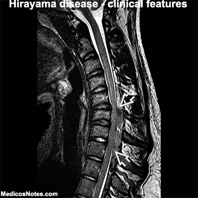
Magnetic Resonance Imaging (MRI): MRI is a key investigation in the diagnosis of Hirayama disease. The imaging findings are typically observed in the cervical spinal cord region, specifically the lower cervical segments (C5-C7). The following MRI findings are characteristic of Hirayama disease:

a. Forward Displacement of the Dural Sac: MRI often reveals a forward displacement of the dural sac during neck flexion. This occurs due to dynamic compression and elongation of the lower cervical spinal cord.

b. Posterior Epidural Space Enlargement: Enlargement of the posterior epidural space, located behind the dural sac, is a common finding in Hirayama disease. This can be visualized as a crescent-shaped area of CSF (cerebrospinal fluid) accumulation.

c. Flattening of the Spinal Cord: MRI may show flattening or concavity of the lower cervical spinal cord, particularly on the posterior aspect, during neck flexion. This is caused by compression of the spinal cord against the anterior vertebral column.

d. Vertebral Column Abnormalities: In some cases, MRI may reveal mild structural abnormalities in the vertebral column, such as focal kyphosis or loss of normal cervical lordosis. These abnormalities contribute to the spinal cord compression during neck flexion.

e. Normal Signal Intensity: Despite the clinical weakness and atrophy observed in Hirayama disease, MRI typically shows normal signal intensity of the affected spinal cord segments. This finding distinguishes it from other progressive motor neuron disorders characterized by abnormal signal intensity on MRI.

It is important to note that the MRI findings may be more pronounced during active neck flexion and may appear normal or less significant during neutral or extended neck positions. Thus, performing MRI with neck flexion is crucial to detect the characteristic findings associated with Hirayama disease.

While clinical evaluation and MRI findings are key in the diagnosis of Hirayama disease, additional investigations, such as laboratory tests and genetic studies, may be performed to exclude other conditions and assess for underlying genetic predispositions. The combination of clinical assessment and MRI findings is essential for a definitive diagnosis of Hirayama disease, enabling appropriate management and support for affected individuals.

Hirayama disease - Treatment

Hirayama Disease - Investigation and MRI findings

Hirayama disease - Clinical Features - An Overview

Understanding Hirayama Disease, Ethiopathogenesis

Hirayama disease - Clinical Features - An Overview

Hirayama disease, also known as juvenile nonprogressive cervical amyotrophy or monomelic amyotrophy, is a rare neurological disorder characterized by distinct clinical features. These features are essential for diagnosing and differentiating Hirayama disease from other neuromuscular conditions. 

Explore the clinical features commonly associated with Hirayama disease:

Age of Onset: Hirayama disease typically manifests in young individuals, predominantly males, in their second or third decade of life. The age of onset ranges from 10 to 25 years, with a peak incidence during adolescence.

Muscle Weakness and Atrophy: The hallmark symptom of Hirayama disease is muscle weakness and atrophy, primarily affecting the muscles of the hand and forearm on one side of the body. The weakness usually begins insidiously and progresses slowly over weeks to months. The atrophy may be more noticeable in the muscles on the back of the hand, leading to a "claw-like" appearance.

Asymmetric Involvement: Hirayama disease typically exhibits asymmetric involvement, with one side of the body being more affected than the other. The weakness and atrophy usually start unilaterally and may eventually spread to involve the other side, although to a lesser extent. This asymmetry helps distinguish Hirayama disease from other progressive neuromuscular disorders.

Muscle Cramps and Fasciculations: Some individuals with Hirayama disease may experience muscle cramps, especially during activity. Fasciculations, which are involuntary muscle twitches or contractions, can also be observed in the affected muscles.

Cold Paresis: A characteristic feature of Hirayama disease is the phenomenon of "cold paresis." Patients may notice worsening of muscle weakness and coordination issues in cold environments or during exposure to cold objects. This symptom is believed to be related to the altered blood flow dynamics and ischemia in the affected spinal cord region.

Sensory Function Preservation: Sensory functions are typically spared in Hirayama disease. Patients do not experience significant sensory loss or abnormalities in the affected limbs. This is another important feature that helps distinguish it from other disorders involving both motor and sensory deficits.

Nonprogressive Course: Hirayama disease is known for its nonprogressive nature. Once the symptoms stabilize, which usually occurs within a few years of onset, further progression is rare. However, it is important to note that the initial progression phase may last several months to a few years before stabilization.

Normal Deep Tendon Reflexes: Deep tendon reflexes, such as the biceps and triceps reflexes, are typically preserved or only mildly affected in Hirayama disease. This finding distinguishes it from other motor neuron disorders characterized by hyperactive or absent reflexes.

It is important to consider these clinical features along with additional diagnostic tests, such as electromyography (EMG), nerve conduction studies, and magnetic resonance imaging (MRI), to arrive at an accurate diagnosis of Hirayama disease. Early recognition and appropriate management can help improve the quality of life for individuals affected by this rare neurological disorder.

Hirayama disease - Treatment

Hirayama Disease - Investigation and MRI findings

Hirayama disease - Clinical Features - An Overview

Understanding Hirayama Disease, Ethiopathogenesis

Understanding Hirayama Disease, ethiopathogenesis


Hirayama disease, also known as juvenile nonprogressive cervical amyotrophy or monomelic amyotrophy, is a rare neurological disorder characterized by muscle weakness and wasting, primarily affecting the muscles of the hand and forearm. The ethiopathogenesis, or the causes and mechanisms, of Hirayama disease have been the subject of scientific investigation, although the exact etiology remains partially understood.

Understanding Hirayama Disease:

Hirayama disease typically affects young individuals, predominantly males, in their second or third decade of life. The condition is more commonly observed in Asian populations, particularly in Japan and India. The clinical course of Hirayama disease is usually nonprogressive, meaning the symptoms do not worsen over time.

Ethiopathogenesis of Hirayama Disease:

Dynamic Compression of the Lower Cervical Spinal Cord: One of the primary theories regarding the ethiopathogenesis of Hirayama disease is dynamic compression of the lower cervical spinal cord. During neck flexion, there is a shift of the posterior dural sac, resulting in anterior displacement and compression of the spinal cord. This compression leads to ischemia and damage to the anterior horns of the spinal cord, causing muscle weakness and atrophy.

Circulatory Disturbances and Vascular Factors: Vascular factors have also been implicated in the development of Hirayama disease. It is believed that the abnormal posture adopted by individuals with this condition, which involves neck flexion and the chin tucking inwards, leads to mechanical compression of the vertebral arteries and subsequent disturbances in blood flow. This compromised blood supply can result in ischemia and neuronal damage in the lower cervical spinal cord.

Genetic Predisposition: Genetic factors may contribute to the development of Hirayama disease. Certain genetic variations and human leukocyte antigen (HLA) associations have been identified in affected individuals, suggesting a potential genetic predisposition. For instance, the HLA-DRB11501 and HLA-DRB11502 haplotypes have been found to be more prevalent in Hirayama disease patients. These genetic factors may influence the susceptibility to spinal cord compression and vascular disturbances.

Immunological Factors: Some studies have proposed an immunological component in the ethiopathogenesis of Hirayama disease. Autoantibodies against various neuronal antigens, such as heat shock proteins, have been detected in the serum and cerebrospinal fluid of affected individuals. These autoantibodies may trigger an autoimmune response that contributes to the damage observed in the lower cervical spinal cord.

Hirayama disease is a rare neurological disorder characterized by muscle weakness and wasting, primarily affecting the muscles of the hand and forearm. The ethiopathogenesis of this condition involves dynamic compression of the lower cervical spinal cord, circulatory disturbances and vascular factors, genetic predisposition, and potential immunological factors. While significant progress has been made in understanding the underlying causes and mechanisms of Hirayama disease, further research is necessary to unravel the complex interplay of these factors and develop more targeted diagnostic and therapeutic strategies for affected individuals.

Hirayama disease - Treatment

Hirayama Disease - Investigation and MRI findings

Hirayama disease - Clinical Features - An Overview

Understanding Hirayama Disease, Ethiopathogenesis


Aorto-Pulmonary Collaterals in Cyanotic Heart Disease: Understanding the Lifelines of Oxygenation


Cyanotic heart disease refers to a group of congenital heart defects characterized by a bluish discoloration of the skin, lips, and nails due to inadequate oxygenation of the blood. One of the significant pathophysiological features of cyanotic heart disease is the presence of aorto-pulmonary collaterals, which play a crucial role in ensuring adequate blood supply to the lungs. In this article, we will explore the importance of aorto-pulmonary collaterals in cyanotic heart disease, their clinical implications, and the strategies used for their management.

Understanding Aorto-Pulmonary Collaterals:

Aorto-pulmonary collaterals are abnormal blood vessels that develop as compensatory pathways between the systemic and pulmonary circulations in individuals with cyanotic heart disease. These collaterals typically arise from the descending thoracic aorta or its branches and supply blood directly to the pulmonary arteries, bypassing the underdeveloped or obstructed native pulmonary circulation. The formation of aorto-pulmonary collaterals is the body's attempt to improve oxygenation by increasing blood flow to the lungs.

Clinical Implications:

The presence of aorto-pulmonary collaterals has several clinical implications in individuals with cyanotic heart disease. Firstly, these collaterals help in maintaining pulmonary blood flow, ensuring that the lungs receive an adequate oxygen supply. Secondly, the collateral vessels can prevent or alleviate the severity of cyanotic spells, which are episodes of acute oxygen desaturation that can lead to cyanosis, breathlessness, and even loss of consciousness. Additionally, aorto-pulmonary collaterals can contribute to the development of pulmonary hypertension, a condition characterized by increased blood pressure within the pulmonary arteries, which can further complicate the management of cyanotic heart disease.

Diagnostic Evaluation:

The evaluation of aorto-pulmonary collaterals in cyanotic heart disease involves a comprehensive diagnostic workup. Non-invasive imaging modalities such as echocardiography, magnetic resonance imaging (MRI), or computed tomography (CT) angiography can provide valuable information about the anatomy, size, and blood flow characteristics of these collaterals. Cardiac catheterization may also be necessary to obtain precise hemodynamic measurements and assess the feasibility of interventional procedures.

Management Strategies:

The management of aorto-pulmonary collaterals in cyanotic heart disease depends on various factors, including the underlying heart defect, the severity of cyanosis, and the presence of associated complications. In some cases, surgical intervention may be required to ligate or disconnect the collaterals to redirect blood flow through the native pulmonary circulation. Alternatively, transcatheter techniques such as coil embolization, occlusion devices, or stent placement can be employed to occlude or restrict the blood flow through the collaterals. The choice of intervention depends on individual patient characteristics and the expertise of the treating team.

Long-Term Follow-Up:

Following the management of aorto-pulmonary collaterals, long-term follow-up is crucial to monitor the effectiveness of the intervention and to detect any potential complications. Regular assessments of oxygen saturation levels, echocardiography, and clinical evaluation are necessary to ensure adequate oxygenation, assess the growth and development of the pulmonary arteries, and identify any signs of recurrent collaterals or other associated anomalies.

Aorto-pulmonary collaterals play a significant role in the pathophysiology of cyanotic heart disease, contributing to the maintenance of pulmonary blood flow and oxygenation. Understanding their significance and effectively managing them is essential for optimizing the outcomes of individuals with cyanotic heart disease. 

Role of Nitric Oxide in Cellular Pathophysiology and Diseases


Nitric oxide (NO) is a small, gaseous signaling molecule with a short half-life, playing crucial roles in various physiological and pathological processes. It is synthesized from L-arginine by the enzyme nitric oxide synthase (NOS) and is involved in several cellular functions, including vasodilation, neurotransmission, and immune regulation. In this article, we will explore the role of nitric oxide in cellular pathophysiology and its association with various diseases.

I. Cellular Pathophysiology of Nitric Oxide

A. Physiological roles of nitric oxide

Vasodilation: NO is a potent vasodilator that helps regulate blood flow and pressure. It stimulates soluble guanylyl cyclase (sGC) in smooth muscle cells, leading to an increase in cyclic guanosine monophosphate (cGMP) levels and subsequent relaxation of the smooth muscle.

Neuronal signaling: NO functions as a neurotransmitter in both the central and peripheral nervous systems, modulating synaptic plasticity, learning, memory, and pain perception.

Immune regulation: NO plays a role in the innate immune response, acting as a cytotoxic molecule against invading pathogens. Macrophages and other immune cells produce NO, which can directly kill pathogens or modulate other immune cells' function.

B. Pathological roles of nitric oxide

Oxidative stress: High concentrations of NO can react with superoxide radicals, forming peroxynitrite, a highly reactive and toxic molecule that contributes to oxidative stress, cell damage, and inflammation.

Inflammation: NO can exacerbate inflammation by upregulating pro-inflammatory cytokines, promoting leukocyte adhesion to the endothelium, and increasing vascular permeability.

Apoptosis: Excessive NO production can trigger apoptosis or programmed cell death, contributing to tissue damage and disease progression.

II. Nitric Oxide in Diseases

A. Cardiovascular diseases

Atherosclerosis: Endothelial dysfunction and decreased NO bioavailability can lead to impaired vasodilation, increased leukocyte adhesion, and platelet aggregation, promoting the development of atherosclerosis.

Hypertension: Reduced NO production or impaired NO signaling can result in vasoconstriction and increased vascular resistance, contributing to hypertension.

Heart failure: Elevated levels of NO, particularly in the failing myocardium, can contribute to left ventricular dysfunction and worsened heart failure prognosis.

B. Neurological disorders

Alzheimer's disease: Impaired NO production and signaling have been implicated in the development of Alzheimer's disease, contributing to neuroinflammation, neuronal damage, and cognitive decline.

Parkinson's disease: Decreased NO bioavailability in the substantia nigra has been linked to dopaminergic neuronal loss in Parkinson's disease.

Stroke: Excessive NO production during ischemic stroke can exacerbate neuronal injury by promoting oxidative stress, inflammation, and apoptosis.

C. Cancer

Aberrant NO signaling can contribute to tumor progression by promoting angiogenesis, inhibiting apoptosis, and modulating the immune response. However, NO can also exert anti-tumor effects by inducing apoptosis and inhibiting cell proliferation, highlighting the dual role of NO in cancer biology.

D. Autoimmune diseases

In autoimmune diseases such as rheumatoid arthritis and multiple sclerosis, NO plays a role in exacerbating inflammation and tissue damage by promoting leukocyte infiltration, upregulating pro-inflammatory cytokines, and contributing to oxidative stress.

Physical Principles of Rotablation: Understanding the Mechanics and Applications


Rotablation, a term derived from "rotational ablation," is a minimally invasive technique used in the field of interventional cardiology for the treatment of heavily calcified coronary arteries. This method employs a high-speed rotational device to abrade and pulverize calcified plaque, thereby restoring blood flow through the coronary artery. In this article, we delve into the physical principles of rotablation, including the mechanics behind the technique and its applications.

I. Fundamentals of Rotablation

A. The Rotablator Device

The rotablator device comprises three main components: a high-speed turbine, a flexible drive shaft, and a diamond-coated burr. The turbine is connected to a console that controls the rotational speed and provides pressurized air or nitrogen to power the system. The drive shaft, encased in a guidewire-compatible catheter, transmits the rotational motion from the turbine to the burr. The diamond-coated burr, available in different sizes, is the component responsible for abrading the calcified plaque.

B. Physics of Rotablation

Centripetal Force: As the burr rotates at high speeds (up to 200,000 RPM), it generates a centripetal force that maintains contact between the burr and the calcified plaque. The force is directly proportional to the square of the burr's rotational speed and the mass of the burr. This centripetal force enables effective ablation of the calcified plaque without causing excessive trauma to the arterial wall.

Particle Size and Speed: The diamond-coated burr's abrasive action pulverizes the calcified plaque into fine particles. These particles are typically smaller than red blood cells, which allows them to be safely cleared from the bloodstream without causing distal embolization or significant vessel injury.

Eccentricity: The eccentricity of the burr allows it to abrade the calcified plaque selectively. The burr rotates off-center, ensuring that it contacts the calcified plaque while avoiding the surrounding healthy arterial tissue.

II. Clinical Applications of Rotablation

A. Coronary Artery Disease (CAD)

Rotablation is primarily used for the treatment of heavily calcified coronary artery lesions that are challenging to manage with conventional percutaneous coronary interventions (PCIs) such as balloon angioplasty and stenting. Calcified plaques are more resistant to dilation and may lead to suboptimal stent deployment or complications such as stent fracture and restenosis. Rotablation can facilitate stent placement by creating a more compliant lesion and reducing the risk of complications.

B. Peripheral Artery Disease (PAD)

In recent years, rotablation has been explored as a treatment option for heavily calcified peripheral artery lesions. Although the technique is less established in this context, preliminary studies have shown promising results in treating calcified lower extremity arteries, improving blood flow, and alleviating symptoms of claudication and critical limb ischemia.

Rotablation is a valuable tool in the armamentarium of interventional cardiologists for the management of heavily calcified coronary artery lesions. The physical principles underlying the technique, such as centripetal force, particle size and speed, and eccentricity, allow for the effective and selective ablation of calcified plaque while minimizing trauma to the arterial wall. As research continues, rotablation may become more widely adopted for treating peripheral artery disease and other vascular conditions.

The Pathophysiology of Acute Aortic Regurgitation: An In-Depth Exploration

Acute aortic regurgitation (AR) is a sudden and severe cardiac condition characterized by the backflow of blood from the aorta into the left ventricle during diastole, which is the relaxation phase of the cardiac cycle. This backflow results from the inadequate closure of the aortic valve, leading to increased pressure on the left ventricle and numerous complications. This article will provide a detailed overview of the pathophysiology of acute aortic regurgitation, its causes, and potential consequences.

1. Causes of Acute Aortic Regurgitation

Acute AR can be triggered by various factors that compromise the integrity of the aortic valve or the aortic root, including:

a. Infective endocarditis: An infection of the endocardium, the inner lining of the heart, can damage the aortic valve leaflets, resulting in AR. This condition is caused by bacteria, most commonly Staphylococcus aureus or Streptococcus species.

b. Aortic dissection: A tear in the aortic wall can lead to the separation of its layers, causing blood to flow into the false lumen and subsequently compromising the aortic valve's function.

c. Traumatic injury: Blunt chest trauma, such as from a car accident or a fall, can damage the aortic valve or root, leading to acute AR.

d. Iatrogenic causes: Medical interventions like aortic valve surgery or catheter-based procedures can inadvertently result in AR.

2. Hemodynamic Consequences

The sudden onset of acute AR leads to several hemodynamic consequences that can rapidly progress to heart failure:

a. Increased left ventricular end-diastolic volume: As blood flows back into the left ventricle, its end-diastolic volume increases, causing the ventricle to dilate and stretch. This leads to a greater preload, or the amount of blood in the ventricle before contraction.

b. Increased left ventricular end-diastolic pressure: The increased volume in the left ventricle elevates the pressure within the chamber, which can eventually be transmitted back into the left atrium, pulmonary veins, and pulmonary capillaries, causing pulmonary congestion and edema.

c. Decreased forward stroke volume: The regurgitant flow reduces the amount of blood ejected into the aorta during systole, which is the contraction phase of the cardiac cycle. This reduction in forward stroke volume can lead to systemic hypotension and poor tissue perfusion.

d. Compensatory mechanisms: In response to the decreased forward stroke volume, the body activates compensatory mechanisms to maintain blood pressure and tissue perfusion, such as increasing heart rate and activating the renin-angiotensin-aldosterone system (RAAS). However, these mechanisms can further exacerbate the heart's workload and contribute to ventricular dysfunction.

3. Ventricular Remodeling and Dysfunction

In acute AR, the rapid increase in left ventricular volume and pressure can cause structural and functional changes in the heart:

a. Myocardial wall stress: The increased volume and pressure within the left ventricle cause the myocardial walls to experience greater stress, which can lead to myocardial ischemia, or inadequate blood supply to the heart muscle.

b. Left ventricular dysfunction: Over time, the increased workload on the left ventricle can lead to systolic and diastolic dysfunction, impairing the heart's ability to contract and relax effectively.

c. Ventricular arrhythmias: The structural and functional changes in the left ventricle can predispose the heart to ventricular arrhythmias, which are abnormal heart rhythms that can be life-threatening.

4. Progression to Heart Failure

The combined effects of increased ventricular volume, pressure overload, and ventricular dysfunction contribute to the development of heart failure in acute AR:

a. Acute left-sided heart failure: The elevated left ventricular pressure and impaired contractility can lead to acute left-sided heart failure, manifesting as pulmonary congestion and edema, shortness of breath, and reduced exercise capacity.

b. Cardiogenic shock: In severe cases, the decreased forward stroke volume and systemic hypotension can result in cardiogenic shock, a life-threatening condition characterized by inadequate tissue perfusion and multi-organ failure.

c. Right-sided heart failure: Prolonged pulmonary congestion and increased pulmonary vascular resistance can eventually lead to right ventricular dysfunction and right-sided heart failure, characterized by peripheral edema, jugular venous distension, and hepatic congestion.

5. Clinical Presentation and Diagnosis

Patients with acute AR may present with a variety of signs and symptoms, such as:

a. Dyspnea: Shortness of breath is a common symptom of acute AR, resulting from pulmonary congestion and reduced oxygen delivery to the tissues.

b. Chest pain: Acute AR can cause chest pain due to myocardial ischemia, increased myocardial oxygen demand, or aortic dissection.

c. Hypotension: Decreased forward stroke volume and systemic hypotension can manifest as dizziness, syncope, and cold extremities.

To diagnose acute AR, clinicians may employ various diagnostic tools, including:

a. Physical examination: A characteristic high-pitched, blowing diastolic murmur can be auscultated at the left sternal border in patients with acute AR.

b. Echocardiography: Transthoracic or transesophageal echocardiography is the gold standard for diagnosing and assessing the severity of AR, providing information on valve morphology, regurgitant flow, and ventricular function.

c. Cardiac catheterization: In some cases, cardiac catheterization may be necessary to confirm the diagnosis, assess coronary artery disease, and evaluate hemodynamics.

Acute aortic regurgitation is a critical cardiac condition that requires prompt diagnosis and management to prevent potentially life-threatening complications, such as heart failure and cardiogenic shock. Understanding the pathophysiology of acute AR enables clinicians to effectively recognize the condition, assess its severity, and initiate appropriate treatment strategies, which may include medical therapy, surgical intervention, or transcatheter valve replacement.

The Deleterious Health Effects of Partially Hydrogenated Vegetable Oils

Partially hydrogenated vegetable oils (PHVOs) are formed when vegetable oils undergo a chemical process called partial hydrogenation. This process adds hydrogen atoms to unsaturated fatty acids in the oil, making it more stable and solid at room temperature. PHVOs are widely used in the food industry due to their longer shelf life, texture-enhancing properties, and heat stability. However, numerous studies have linked PHVOs to several health issues, primarily due to the formation of trans fatty acids (TFAs) during the hydrogenation process. This article examines the deleterious health effects associated with PHVO consumption and the importance of seeking healthier alternatives.

Cardiovascular Diseases

The consumption of PHVOs has been strongly linked to an increased risk of cardiovascular diseases (CVDs). TFAs can raise low-density lipoprotein (LDL) cholesterol levels, also known as "bad" cholesterol, while simultaneously lowering high-density lipoprotein (HDL) cholesterol or "good" cholesterol. This imbalance contributes to the build-up of plaque in the arteries, which can eventually lead to heart attacks, strokes, and other cardiovascular complications. A meta-analysis of several studies revealed that a 2% increase in energy intake from TFAs corresponds to a 23% increase in the risk of coronary heart disease.

Inflammation and Endothelial Dysfunction

PHVO consumption has also been associated with increased inflammation and endothelial dysfunction, both of which are critical factors in the development of atherosclerosis. TFAs are known to trigger inflammatory responses by activating pro-inflammatory cytokines and increasing C-reactive protein (CRP) levels. In addition, they can impair the functioning of the endothelium, the inner lining of blood vessels, by reducing the bioavailability of nitric oxide, a vasodilator essential for maintaining vascular health.

Type 2 Diabetes

A diet high in PHVOs may contribute to the development of type 2 diabetes by negatively affecting insulin sensitivity. In several observational studies, higher TFA intake has been linked to a higher risk of type 2 diabetes. The mechanism underlying this relationship is not entirely understood, but it is suggested that TFAs may alter cell membrane structure and function, disrupt insulin signaling, and promote inflammation, which can impair glucose metabolism.

Obesity

Although the relationship between PHVO consumption and obesity remains inconclusive, some studies have suggested that TFAs may contribute to weight gain and abdominal fat accumulation. A potential explanation for this effect is that TFAs may disrupt the activity of key enzymes involved in lipid metabolism, such as lipoprotein lipase and hormone-sensitive lipase, which could lead to increased fat storage.

Cancer

Some research has indicated that high TFA intake may be associated with an increased risk of certain cancers, such as breast, colon, and prostate cancer. While the exact mechanisms are not yet fully understood, it is hypothesized that TFAs may promote inflammation, disrupt cell signaling pathways, and interfere with the normal functioning of cell membranes, which can contribute to the development and progression of cancer.

The deleterious health effects of PHVOs can no longer be ignored. Due to the extensive evidence linking their consumption to a wide range of health issues, many countries have implemented policies to reduce or eliminate TFAs from the food supply. As consumers, it is essential to be aware of the risks associated with PHVO consumption and opt for healthier alternatives, such as unhydrogenated vegetable oils, which contain fewer TFAs and have been associated with more favorable health outcomes.

The Anatomy of the Mitral Apparatus: Structure and Function


The mitral apparatus is a complex structure located within the left side of the heart, responsible for ensuring the efficient unidirectional flow of blood from the left atrium to the left ventricle. A thorough understanding of the mitral apparatus's anatomy is crucial for healthcare professionals, especially those in cardiology, to diagnose and treat related disorders effectively. This article will discuss the various components of the mitral apparatus, their individual functions, and their collective role in maintaining proper cardiac function.

I. Components of the Mitral Apparatus

The mitral apparatus comprises several interconnected structures that work in harmony to ensure the proper functioning of the mitral valve. These components include:

  1. Mitral valve leaflets
  2. Annulus fibrosus
  3. Chordae tendineae
  4. Papillary muscles

II. Mitral Valve Leaflets

The mitral valve consists of two leaflets, the anterior and the posterior leaflet, that form a seal between the left atrium and the left ventricle. The anterior leaflet, also known as the aortic leaflet, is larger and more crescent-shaped, while the posterior leaflet, or mural leaflet, is smaller and more quadrangular. Both leaflets are composed of three layers: the atrialis, the spongiosa, and the ventricularis. During ventricular systole, the leaflets coapt and prevent the backflow of blood into the left atrium.

III. Annulus Fibrosus

The annulus fibrosus is the fibrous ring that forms the base of the mitral valve, anchoring it to the left atrium and left ventricle. The annulus fibrosus is not a perfect circle but has a saddle-shaped, dynamic structure that undergoes changes throughout the cardiac cycle. It provides support and stability to the mitral valve leaflets and helps maintain their proper alignment.

IV. Chordae Tendineae

The chordae tendineae are thin, fibrous cords that connect the mitral valve leaflets to the papillary muscles. They are primarily composed of collagen and elastin fibers and are responsible for preventing the prolapse of the mitral valve leaflets into the left atrium during ventricular systole. The chordae tendineae are classified into three types based on their attachment points on the mitral leaflets:

Primary chordae: Also known as marginal chordae, they attach directly to the free edge of the mitral leaflets.

Secondary chordae: Also known as intermediate chordae, they connect to the ventricular surface of the mitral leaflets.

Tertiary chordae: Also known as basal chordae, they attach to the atrial surface of the mitral leaflets.

V. Papillary Muscles

The papillary muscles are cone-shaped muscular projections from the left ventricular wall that serve as the anchor points for the chordae tendineae. There are two main papillary muscles in the left ventricle: the anterolateral and the posteromedial papillary muscles. These muscles contract during ventricular systole, pulling on the chordae tendineae to keep the mitral valve leaflets in a closed position and preventing mitral regurgitation.

VI. The Role of the Mitral Apparatus in Cardiac Function

The mitral apparatus plays a critical role in maintaining efficient cardiac function by ensuring the unidirectional flow of blood from the left atrium to the left ventricle. During diastole, the mitral valve opens, allowing blood to flow from the left atrium to the left ventricle. As the ventricle contracts during systole, the mitral valve closes, preventing the backflow of blood into the left atrium. The coordinated action of the mitral valve leaflets, annulus fibrosus, chordae tendineae, and papillary muscles ensures the proper functioning of the mitral apparatus throughout the cardiac cycle.

VII. Clinical Significance

A thorough understanding of the mitral apparatus's anatomy is essential for healthcare professionals to diagnose and treat disorders related to the mitral valve effectively. Common conditions affecting the mitral apparatus include mitral valve prolapse, mitral regurgitation, and mitral stenosis. These conditions can lead to impaired cardiac function, heart failure, and other serious complications if left untreated.

Modern diagnostic techniques, such as echocardiography, cardiac magnetic resonance imaging (MRI), and computed tomography (CT), can help visualize the mitral apparatus's structure and function in detail. This information is crucial for planning interventions, such as mitral valve repair or replacement surgery, to restore normal cardiac function.

The mitral apparatus is a complex and vital structure within the heart that ensures the unidirectional flow of blood from the left atrium to the left ventricle. Its components, including the mitral valve leaflets, annulus fibrosus, chordae tendineae, and papillary muscles, work in harmony to maintain proper cardiac function. A comprehensive understanding of the mitral apparatus's anatomy is crucial for healthcare professionals to diagnose and treat related disorders effectively and improve patient outcomes.

The Fetal Circulatory Changes at Birth: A Cardiology Perspective


The fetal circulatory system undergoes significant changes during birth as the newborn transitions from a prenatal to a postnatal environment. Understanding these changes is crucial for healthcare professionals, particularly those in the field of cardiology, to ensure the healthy development of the newborn. This article will provide an overview of the fetal circulatory system, the key changes that occur at birth, and the importance of these changes for the newborn's survival and well-being.

I. The Fetal Circulatory System

During pregnancy, the fetus receives oxygen and nutrients from the mother's placenta via the umbilical cord. The fetal circulatory system is adapted to this unique environment, allowing the blood to bypass the non-functional fetal lungs and liver.

Key features of the fetal circulatory system include:

The placenta: The primary organ of gas exchange, nutrient supply, and waste removal between the mother and the fetus.

The umbilical vein: Carries oxygenated blood from the placenta to the fetus.

Ductus venosus: A shunt that diverts a portion of the oxygenated blood from the umbilical vein away from the liver and directly into the inferior vena cava.

Foramen ovale: An opening in the atrial septum that allows oxygenated blood to flow from the right atrium to the left atrium, bypassing the lungs.

Ductus arteriosus: A short vessel that connects the pulmonary artery to the aorta, diverting blood away from the fetal lungs.

II. Circulatory Changes at Birth

At birth, the newborn starts to breathe, and the placental circulation is severed. This leads to a series of physiological changes in the circulatory system to establish the postnatal circulation:

Closure of the umbilical vessels: With the severing of the umbilical cord, the umbilical vein and arteries close, ending the blood flow between the placenta and the newborn.

Increased systemic resistance: Due to the loss of the low-resistance placental circulation, systemic vascular resistance increases, leading to a rise in blood pressure.

Initiation of pulmonary circulation: The newborn's first breaths cause the lungs to expand and fill with air, reducing pulmonary vascular resistance and increasing pulmonary blood flow.

Closure of the foramen ovale: Increased blood return to the left atrium and decreased pressure in the right atrium result in the functional closure of the foramen ovale, which eventually becomes the fossa ovalis.

Closure of the ductus arteriosus: Increased oxygen concentration in the blood and decreased prostaglandin levels lead to the constriction and eventual closure of the ductus arteriosus, typically within the first few days of life.

Closure of the ductus venosus: The ductus venosus constricts and closes due to increased hepatic blood flow and decreased prostaglandin levels, eventually becoming the ligamentum venosum.

III. Significance of Circulatory Changes

The circulatory changes at birth are vital for the newborn's survival and adaptation to life outside the womb. These changes ensure that the blood flow is redirected from the non-functional fetal organs to the fully functional organs in the newborn's body, facilitating proper growth and development.

The fetal circulatory changes at birth are crucial for the transition from a prenatal to a postnatal environment. Understanding these changes is essential for healthcare professionals, particularly in cardiology, to monitor and care for newborns effectively. Early detection and management of any abnormalities in the circulatory system can significantly impact a newborn's long-term health and development.